40 diagram of uterus and bladder
Prolapse of the Uterus, Bladder, Bowel, or Rectum. If you have questions or need a physician referral, please contact HERS at 610-667-7757. Broad bands of uterine ligaments provide structural support to the uterus and pelvis. The uterine ligaments may weaken, stretch, or they can be damaged or severed during surgery. Positions of uterus. Diagram for variants of uterine position. Normal uterus rests on the superior surface of the empty bladder. Normal Uterus Positions. Artistic style female reproductive system vector illustration educational poster. Health and medicine labeled diagram. Female sexual organ cross section with
Computer illustration of the female reproductive system (pink) and bladder (yellow). The endometrium (red) is extending outside the uterus, into the fallopian tube, ovaries and abdominal cavity. This condition is known as endometriosis, and causes intense pain in the pelvic region.

Diagram of uterus and bladder
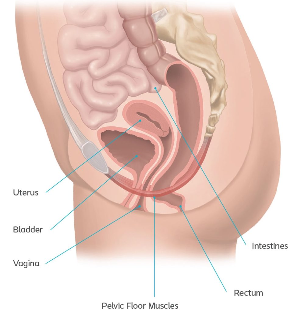
Uterine prolapse occurs when pelvic floor muscles and ligaments stretch and weaken and no longer provide enough support for the uterus. As a result, the uterus slips down into or protrudes out of the vagina. Uterine prolapse can occur in women of any age. But it often affects postmenopausal women who've had one or more vaginal deliveries. Browse 213 uterus diagram stock photos and images available, or search for female reproductive system or uterus icon to find more great stock photos and pictures. An anatomical diagram depicts the method of extracting a fetus by reaching into the uterus and adjusting the baby's position. Located underneath the pelvis, the pelvic floor not only supports the uterus and vagina, but also the bladder, intestines and rectum - ultimately holding them in place and allowing them to function correctly. It is these muscles that will play a major part in the use and removal of your menstrual cup.
Diagram of uterus and bladder. At this point, your uterus has begun to grow and become more egg-shaped. The pressure of the growing uterus on the bladder causes frequent urge to urinate. In this image, you can see the beginnings of the placenta in the uterus. The embryo is about 1/4 inch to 1/2 inch long and weighs 1/1,000th of an ounce. The uterus, also known as the womb, is the hollow, pear-shaped organ in the female pelvis in which fertilization of an ovary (egg), implantation of the resulting embryo, and development of a baby take place. ... Shaped like an inverted pear, the uterus sits behind the bladder and in front of the rectum. The uterus sits in the center of the pelvis held in place by four sets of ligaments. The uterus separates the bladder and the bowel and holds those organs in their rightful positions. Once the ligaments are severed and the uterus removed, the bladder and bowel drop down and, without the uterus to separate them, are now adjacent to each other. The empty urinary bladder is somewhat tetrahedral in shape - like a three sided pyramid with a triangular base (illustrated in diagram). This gives the bladder one superior surface (top), two inferolateral surfaces (sides) and a posterior surface (back). The external aspect of the superior surface of the bladder is covered by peritoneum.
The urinary bladder is found inferior to the peritoneum, sitting on the pelvic floor. In females its inferior surface lays on the pubic symphysis and the posterior wall is in contact with the vagina and uterus.. In males, the inferior surface of the bladder lays over the pubic symphysis and prostate, posteriorly is the distal third of the rectum.Between the posterior surface of the bladder and ... Start studying Female Urinary Bladder and Urethra. Learn vocabulary, terms, and more with flashcards, games, and other study tools. Below and in front of the uterus is the bladder. The bladder, also known as the urinary bladder, is an expandable, muscular sac that stores urine. When signaled, ... The female pelvic organs: bladder, vagina, uterus, fallopian tube, ovaries. Location and overview
Position. The normal position of uterus between the bladder and the rectum, and it projects superoanteriorly over the urinary bladder. Normally and most commonly it is anteverted and anteflexed. The long axis of vagina forms a ninety degree angle with axis of cervix which is directed posteriorly and inferiorly, this is called as anteversion. The urinary bladder stores urine prior to its elimination from the body (functions of the urinary system). At micturation / urination, the bladder expels urine into the urethra, leading to the exterior of the body. The bladder is a musculomembranous sac located on the floor of the pelvic cavity, anterior to the uterus and upper vagina (in females). Anatomy. The bladder is a triangle-shaped, hollow organ. In men, it is bordered by the pubic bone at the front of the pelvis and the rectum at the back of the pelvis in the lower abdomen. In women, the bladder is bordered posteriorly by the uterus and vagina. 1 The bladder is supported by ligaments and connects at the top to two ureters ... Browse 878 uterus diagram stock illustrations and vector graphics available royalty-free, or search for female reproductive system or uterus icon to find more great stock images and vector art. Newest results. female reproductive system. uterus icon.
Anatomy location of bladder, vaginal canal, cervix and ...
The uterus is also shown. Anatomy of the female urinary system showing the kidneys, ureters, bladder, and urethra. Urine is made in the renal tubules and collects in the renal pelvis of each kidney. The urine flows from the kidneys through the ureters to the bladder. The urine is stored in the bladder until it leaves the body through the urethra.
Stomach, liver, intestine, bladder, lung, testicle, uterus, spine, pancreas, kidney, heart, bladder icon. Donor medical poster diagram of body organs female pics stock illustrations. Brain Power - Businesswoman Businesswoman working on a brain diagram of body organs female pics stock illustrations.
Uterus: Anatomy, blood supply, histology, functions | Kenhub
The female urogenital tract consists of all the organs involved in reproduction and the formation and release of urine. It includes the kidneys, ureters, bladder, urethra, and the organs of reproduction - uterus, ovaries, fallopian tubes and vagina. The kidneys are bean shaped organs, which help the body produce urine to get rid of unwanted ...
Factors commonly associated with causing a prolapsed bladder are those that weaken the pelvic floor muscles and ligaments that support the bladder, urethra, uterus, and rectum, which can lead to detachment from the ligaments or pelvic bone where the muscles attach: Pregnancy and childbirth: This is the most common cause of a prolapsed bladder.

Female reproductive organs. a) non-gravid uterus (mice and ...
The bladder, like the stomach, is an expandable saclike organ that contracts when it is empty. The inner lining of the bladder tucks into the folds and expands out to accommodate liquid. When ...

Diagram illustrating the uterus after delivery of the ...
This medical exhibit diagram illustrates the anatomy of the female abdomen and pelvis from an anterior (front) cut-away view, showing elements of the digestive system. The liver, stomach, and abdominal contents are clearly identified and labeled, including the cecum, ascending colon, transverse colon, descending colon, and small intestine. The image also shows the pelvis, uterus, and urinary ...

Diagram Of Uterus And Bladder - Hanenhuusholli
In females, the bladder lies inferior to uterus and ahead to the vagina. The size of the bladder is about the shape of a pear when empty, its normal capacity is 400ml to 600ml. Prolapsed Bladder or also called Cystocele is a condition when the muscle between women's bladder and vagina fall out of place, and sticks out into vagina.
NORMAL ANATOMY OF THE FEMALE PELVIS AND TRANSVAGINAL ...
The uterus and the bladder are held in their normal positions just above the inside end of the vagina by a "hammock" made up of supportive muscles and ligaments. Wear and tear on these supportive structures in the pelvis can allow the bottom of the uterus, the floor of the bladder or both to sag through the muscle and ligament layers.

Image from page 1417 of "Anatomy, descriptive and applied" (1913)
Diagram of uterus and bladder. The bladder is connected to the kidneys by two long tubes called ureters. When empty the bladder is about the size and shape of a pear. On the other hand the bladder exhibits an oval shape when it is full. When this occurs the uterus or bladder can create a bulge into the vagina.

Female Pelvis | Radiology Key
The diagram represents a sagittal view of the uterus reflecting an 'L" shaped structure of the uterus vagina, and the internal cavity. The parts that the tract traverses includes the vagina at the downstream end which now is a significantly (pink), cervix (royal blue), lower uterine segment (light blue), the body (lighter blue), and the ...

Image from page 357 of "Comparative anatomy of vertebrates" (1912)
The pelvic cavity is a bowl-like structure that sits below the abdominal cavity. The true pelvis, or lesser pelvis, lies below the pelvic brim (Figure 1). This landmark begins at the level of the sacral promontory posteriorly and the pubic symphysis anteriorly. The space below contains the bladder, rectum, and part of the descending colon. In females, the pelvis also houses the uterus ...

(A) Stage 2 anterior compartment prolapse. (B) Cervix on ...
Located underneath the pelvis, the pelvic floor not only supports the uterus and vagina, but also the bladder, intestines and rectum - ultimately holding them in place and allowing them to function correctly. It is these muscles that will play a major part in the use and removal of your menstrual cup.

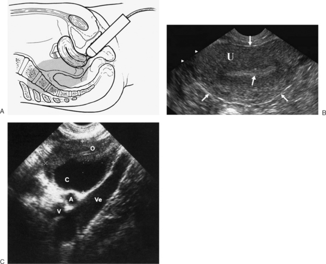
Transabdominal ultrasound of the ovary. Identify urinary ...
Browse 213 uterus diagram stock photos and images available, or search for female reproductive system or uterus icon to find more great stock photos and pictures. An anatomical diagram depicts the method of extracting a fetus by reaching into the uterus and adjusting the baby's position.

Image from page 246 of "The hydropathic encyclopedia : a system of hydropathy and hygiene in eight parts ... designed as a guide to families and students, and a text-book for physicians" (1853)
Uterine prolapse occurs when pelvic floor muscles and ligaments stretch and weaken and no longer provide enough support for the uterus. As a result, the uterus slips down into or protrudes out of the vagina. Uterine prolapse can occur in women of any age. But it often affects postmenopausal women who've had one or more vaginal deliveries.

Image from page 758 of "The cyclopædia of anatomy and physiology" (1859)

Anatomy of female uterus with ovaries kidney and bladder ...
Anatomical maps of the female pelvis at the level of the ...

Image from page 471 of "Treatise on gynaecology : medical and surgical" (1894)

John Yesko: Chicago user experience design, information ...

Pelvic ultrasonography showing in A -normal cervix, uterus ...

Anatomy of the female pelvis with relations of the urinary ...

Duplicated uterus and two hemi-vagina with ectopic ureter ...

32 Diagram Of Uterus And Bladder - Wiring Diagram List

Perineal trauma dissertation titles

Image from page 71 of "Manual of gynecology" (1883)

32 Diagram Of Uterus And Bladder - Wire Diagram Source ...

Elevation Physiotherapy | Pelvic Organ Prolapse—What Can ...

Image from page 245 of "A laboratory manual and text-book of embryology" (1915)
Clinical Reproductive Anatomy - Uterus - 3D Anatomy ...

Transabdominal ultrasound of the uterus. Note the urinary ...

Image from page 512 of "Clinical gyncology, medical and surgical" (1895)

Diagram Uterus Bladder
Female Pelvis and Female Reproductive System - Beata's DMS ...
The womb - Understanding - Macmillan Cancer Support
Learn More About Pelvic Organ Prolapse Symptoms & Treatments
Pelvis Anatomy Illustrations for Presentations and ...

Pain during sex and chronic pelvic pain | BabyCenter
AmateurCoedSex

Robotic sacrocolpopexy - cystocele & large rectocele - YouTube
0 Response to "40 diagram of uterus and bladder"
Post a Comment