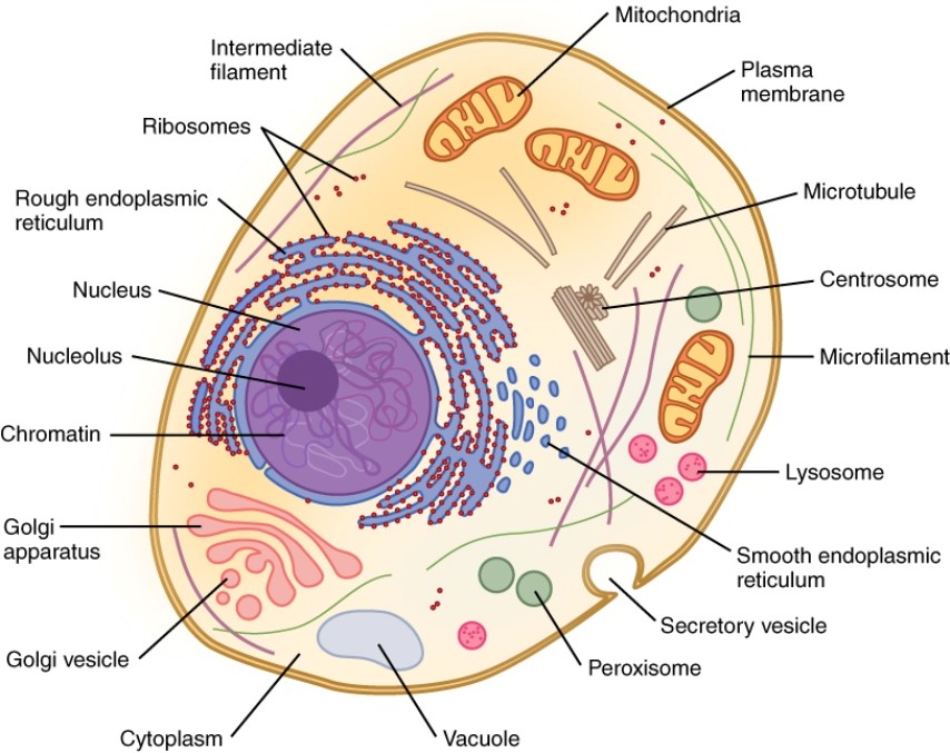
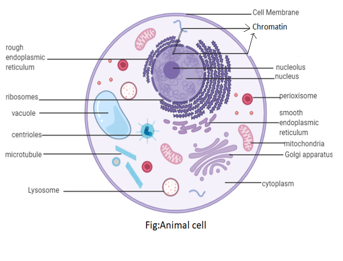
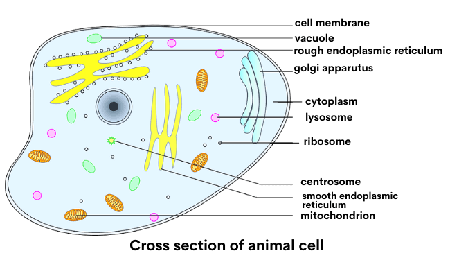
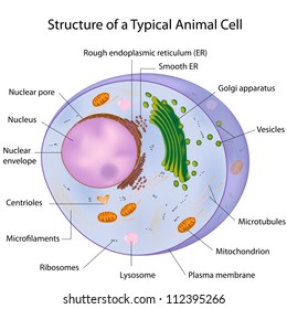
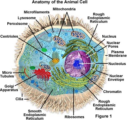
43 animal cells diagram with labels
The animal cell diagram is widely asked in Class 10 and 12 examinations and is beneficial to understand the structure and functions of an animal. A brief explanation of the different parts of an animal cell along with a well-labelled diagram is mentioned below for reference. This packet includes diagrams to label, graphic organizers, and a quiz on plant and animal cells. Download the preview to take a peek at the contents. Table of Contents: p. 1 Cover p. 2 Table of Contents p. 3 Essential Questions p. 4 Facts about cells graphic organizer - Have students fold the oute
Learning about animal cells can be tricky at first, so why don't we start you off relatively easy with this animal cell part labelling quiz! In this one we'll be giving you a question referring to a given diagram and asking you to label it. Simple, right? Let's find out how many you get right.
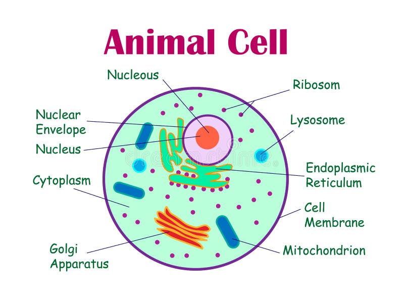
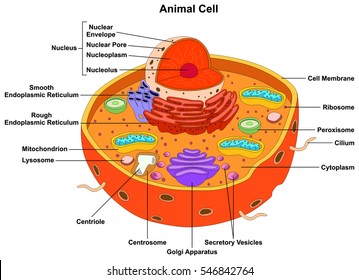
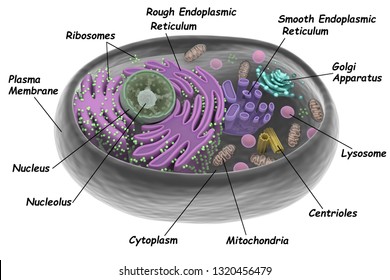
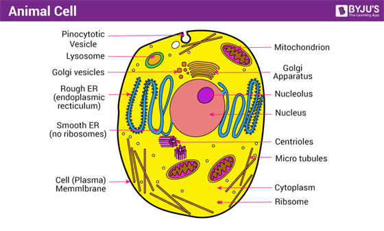
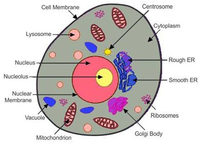
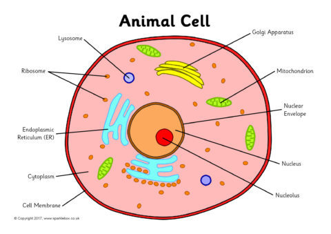
Animal cells diagram with labels
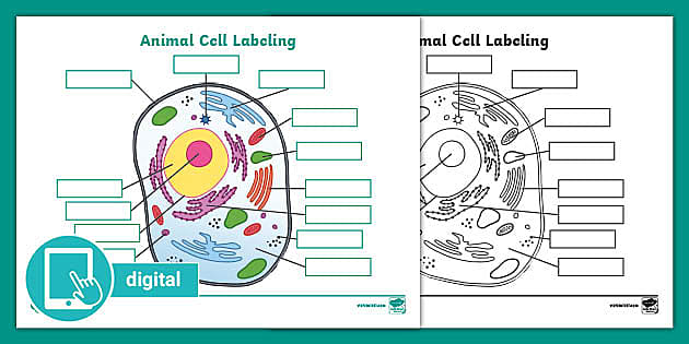
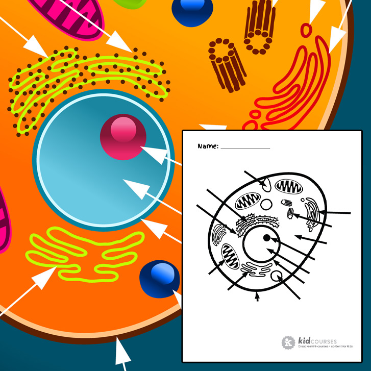
Start studying Animal Cell Labeling. Learn vocabulary, terms, and more with flashcards, games, and other study tools. The Animal Cell Worksheet Name: Label the animal cell drawn below and then give the function of each cell part. (Note: The lysosomes are oval and the vacuoles are more rounded.) 1. 7. 8. 2. 9. 3. 10. 4. 11. 5. 6. Cell Part: Function of Cell Part: 12. nucleus 13. endoplasmic reticulum 14. ribosome 15. cytoplasm 16. nucleolus 17. Mitosis is a process of cell division which results in the production of two daughter cells from a single parent cell. The daughter cells are identical to one another and to the original parent cell. In a typical animal cell, mitosis can be divided into stages: Interphase: ... Labeled Diagram Author: jana stewart Last modified by: jana stewart ...
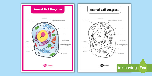
Animal cells diagram with labels. This is a basic illustration of a plant cell with major parts labeled. Labels include nucleus, chloroplast, cytoplasm, membrane, cell wall, and vacuole, and mitochondrion. Use it as a poster in your classroom or have students glue it into their science notebooks. An animal cell diagram is a great way to learn and understand the many functions of an animal cell. The diagram, like the one above, will include labels of the major parts of an animal cell including the cell membrane, nucleus, ribosomes, mitochondria, vesicles, and cytosol. The cells of animals are the basic structural units for the wide ... Animal Coloring Pages. Animal Cell Diagram. Teach your students all about the inner working of an animal cell with the help of this hand-drawn animal cell diagram. This pdf packet contains 6 versions of the diagram, to help you teach and also quiz your students:1. Labeled Animal Cell Diagram - Color2. Answer 5 questions correctly in a row. Label the two cell parts on the diagram below. chromosomes. vacuoles. cell membrane. endoplasmic reticulum. nucleus. Created with Snap. Learn with an example.
Labelled diagrams of typical animal and plant cells with editable layers. The structure of an animal cell, with labeled parts. Biology vector illustration. Start studying Label Plant & Animal Cells. Learn vocabulary, terms, and more with flashcards, games, and other study tools. Learn about the animal cell diagram labeled, parts of an animal cell with their functions, differences & similarities between plant and animal cells, types of an animal cells, and other important facts and information about animal cells. The typical cell ranges in size between 1-100 micrometers. The lack of cell walls enabled the animal cells to develop a greater diversity of cell types. Animals possess a diversity of characters ranging from corals to large carnivores. The lack of cell walls made them develop characters such as defense, flexibility, and reproduction. Diagram of ...
Glossary of Animal Cell Terms: · Cell Membrane: The thin layer of protein and fat that surrounds the cell. · Centrosome (Microtubule Organizing Center) · Cytoplasm ... Stage 1. Answer 4 questions correctly in a row. Label the two cell parts on the diagram below. Plant cells contain many organelles such as ribosomes, the nucleus, the plasma membrane, the cell wall, mitochondria, and chloroplasts. In addition, plant cells differ from animal cells in a number of key ways. Examining a diagram of the plant cell will help make the differences clearer. Let's go over the individual components of plant cells ... The following animal cell diagram labeled show more parts of the cell. Animal cell contains membrane bound nucleus, it also contains other membrane bound cellular organelles. These cellular organelles carry out specific functions that are necessary for the normal functioning of the cell. Animal cells lack cell wall, a large vacuole and plastids.
Animal Cells Are Eukaryotic Cells With a Membrane-Bound Nucleus Animal cells contain membrane-bound organelles (tiny cellular structures) that carry out specific functions necessary for normal cellular operation.
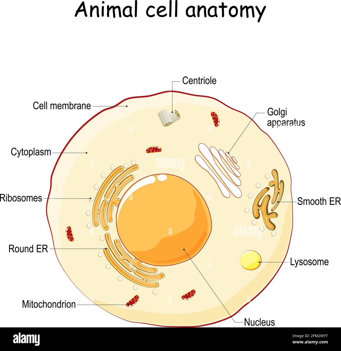
Animal Cell Anatomy Vector Diagram The Structure Of A Human S Cell With Labeled Parts Cross Section Of A Eukaryotic Cell Illustration For Biology Stock Vector Image Art Alamy
Mar 18, 2014 - Can you label and color these important parts of the animal cell? NUCLEUS control center for cell (cell growth, cell metabolism, ...
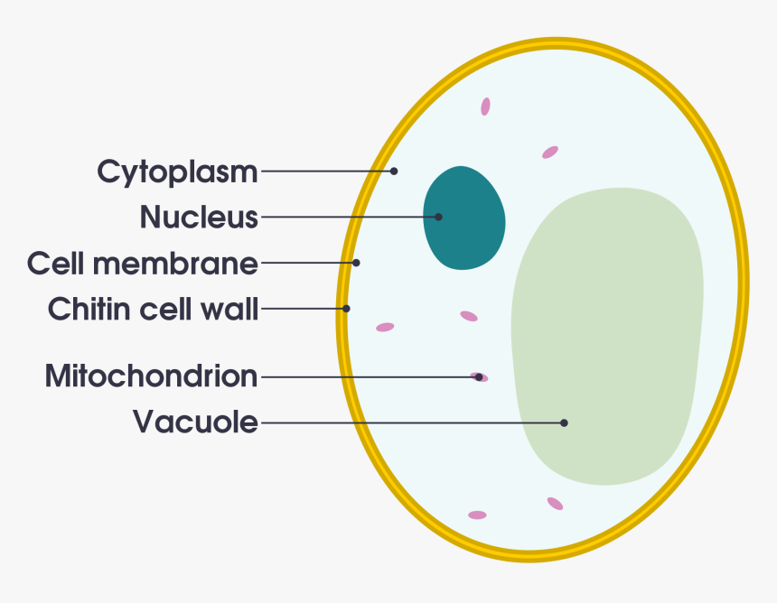
Animal cells and plant cells are similar in that they are both eukaryotic cells. Plant cells have three extra components, a vacuole, chloroplast and a cell wall. A bacteria diagram basically helps us to profit more about this unmarried cell organisms that have neither membrane-bounded nucleolus or organelles like mitochondria and chloroplasts.
Animal Cell Anatomy. The cell is the basic unit of life. All organisms are made up of cells (or in some cases, a single cell). Most cells are very small; in fact, most are invisible without using a microscope. Cells are covered by a cell membrane and come in many different shapes.
Browse 19 animal cell labeled diagram stock photos and images available, or start a new search to explore more stock photos and images. Newest results. Diagrams of animal and plant cells Labelled diagrams of typical animal and plant cells with editable layers. animal cell labeled diagram stock illustrations.
Labeled Animal Cell Diagram diagram and chart - Human body anatomy diagrams and charts with labels. This diagram depicts Labeled Animal Cell Diagram.Human anatomy diagrams show internal organs, cells, systems, conditions, symptoms and sickness information and/or tips for healthy living.
12. $2.00. PDF. Three versions of the plant cell worksheet and three versions of the animal cell worksheet allow students of different grade levels and/or skill levels to label and review the parts of each cell type. Can be used as homework or a quiz. Answer key is provided and can be projected and used during less.
Human Cell Diagram, Parts, Pictures, Structure and Functions The cell is the basic functional in a human meaning that it is a self-contained and fully operational living entity. Humans are multicellular organisms with various different types of cells that work together to sustain life.
Plant and Animal Cell: All the plants and animals are made up of a single unit called a cell. Robert Hooke discovered the cell in the year \(1665\). PPLO is the smallest known cell. The longest cells are the nerve cells.
Investigating Animal Cells Today you will use the link below to visit a website to research animal cells. In your INB, you will need to make sure your diagram and chart are completed. You will be able to find all of this information on the website listed below. Cells Alive! Animal Cell Exploration
Animal Cell - Science Quiz: Animal cells are packed with amazingly specialized structures. One vital part of an animal cell is the nucleus. It's the cell's brain, employing chromosomes to instruct other parts of the cell. The mitochondria are the cell's powerplants, combining chemicals from our food with oxygen to create energy for the cell.
However, the cell membrane in plant cells is quite rigid, while, the cell membrane in animal cells is quite flexible. As observed in the labeled animal cell diagram, the cell membrane forms the confining factor of the cell, that is it envelopes the cell constituents together and gives the cell its shape, form, and existence.
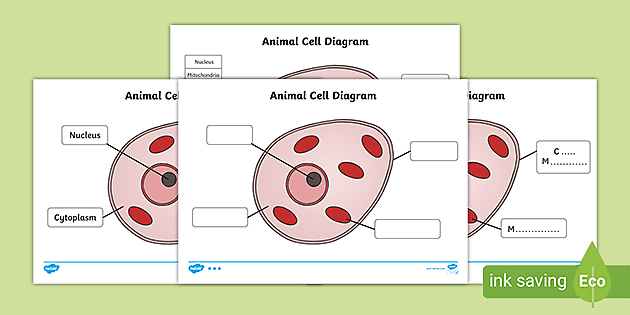
If your students find the Animal Cell Labelling Worksheet useful, this Plant Cell Diagram is a similar labelling activity for plant cells. For something a little more challenging, this Parts of a Cell Cut and Stick Worksheet also challenges students to recall the different components of both animal and plant cells.
by Jordanu176. G2 Science Animal Needs Life Science. label the secret coders Labelled diagram. by Secretcoder. Label Secret coders. April Animal Reproduction and Cell Reproduction Vocab Review, Solimene Match up. by Solims. Cell structure Labelled diagram. by Jacqueline55.
Mitosis is a process of cell division which results in the production of two daughter cells from a single parent cell. The daughter cells are identical to one another and to the original parent cell. In a typical animal cell, mitosis can be divided into stages: Interphase: ... Labeled Diagram Author: jana stewart Last modified by: jana stewart ...
The Animal Cell Worksheet Name: Label the animal cell drawn below and then give the function of each cell part. (Note: The lysosomes are oval and the vacuoles are more rounded.) 1. 7. 8. 2. 9. 3. 10. 4. 11. 5. 6. Cell Part: Function of Cell Part: 12. nucleus 13. endoplasmic reticulum 14. ribosome 15. cytoplasm 16. nucleolus 17.
Start studying Animal Cell Labeling. Learn vocabulary, terms, and more with flashcards, games, and other study tools.

25 Animal Cells Diagram With Labels Markcritz Template Design Cell Diagram Cell Diagram Project Plant And Animal Cells

Biology Diagram Show Structure Of Animal And Plant Cell Stock Illustration Download Image Now Istock












0 Response to "43 animal cells diagram with labels"
Post a Comment