43 sheep brain labeled diagram
Quizlet makes simple learning tools that let you study anything. Start learning today with flashcards, games and learning tools — all for free. to anatomy studies. See for yourself what the . cerebrum, cerebellum, spinal cord, gray matter, white matter, and other parts of the brain look like! Observation: External Anatomy . 1. You'll need a . preserved sheep brain. for the dissection. Set the brain down so the flatter side, with the white . spinal cord. at one end, rests on the ...
April 9, 2018 - Brain Of The Sheep – Labeled in Blank Sheep Brain Diagram Sheep Brain Dissection Guide With Pictures | Secondary Science in Blank Sheep Brain Diagram Sheep Brain Dissection Lab Sheet | Ap Psychology Prep | Pinterest in Blank Sheep Brain Diagram Brain Dissection Diagram Sheep Brain Anatomy ...

Sheep brain labeled diagram
DISSECTION OF THE SHEEP'S BRAIN Introduction The purpose of the sheep brain dissection is to familiarize you with the three-dimensional structure of the brain and teach you one of the great methods of studying the brain: looking at its structure. One of the great truths of studying biology is the saying that "anatomy precedes physiology". This is an online quiz called Sheep Brain. This quiz has tags. Click on the tags below to find other quizzes on the same subject. Anatomy. sheep brain. Labeled Sheep Heart Picture #1. Explore nicellian's photos on Flickr. nicellian has uploaded 157 photos to Flickr.
Sheep brain labeled diagram. Psychology . Department · Murray Research Center Want more on Memory? Visit our · Comments? Contact: Webteam © 1998-1999 Exploratorium Sheep Brain Dissection with Labeled Images The sheep brain is exposed and each of the structures are labeled and described in a sequential manner, in the same way that a real dissection would occur. Amanda Huss Dissection guide with instructions for dissecting a sheep brain. Checkboxes are used to keep track of progress and each structure that can be found is described with its location in relation to other structures. An image of the brain is included to help students find the structures.
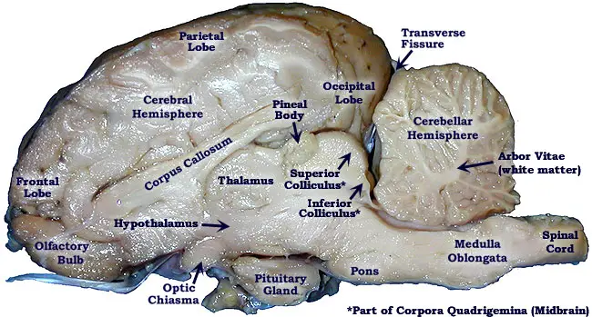
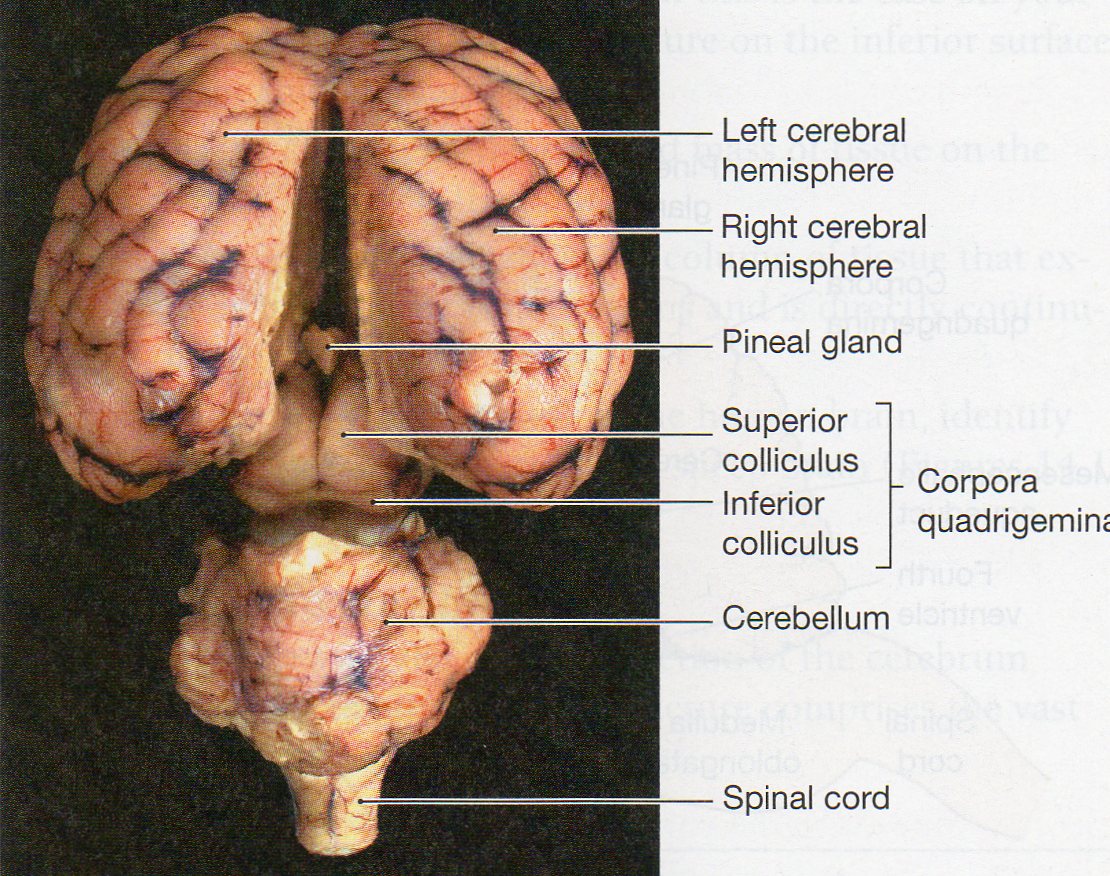
Carolina's Perfect Solution® sheep brain dissection introduces students to the anatomy of a mammalian brain, both external and internal, and encourages students to construct an explanation of the central nervous system. Below is a brief survey of the internal and external anatomy of the sheep brain. Diagram Worksheets. Label the Parts of a Sheep Brain. Print out these diagrams and fill in the labels to test your knowledge of sheep brain anatomy. Internal anatomy: label the right side (.pdf) External anatomy: label the top view (.pdf) External anatomy: label the bottom view (.pdf) What other users say: Fun and Educational. Start studying Sheep Brain Dissection labeled 2. Learn vocabulary, terms, and more with flashcards, games, and other study tools. The lobes of the brain are visible, as well as the transverse fissure, which separates the cerebrum from the cerebellum. The convolutions of the brain are also visible as bumps (gyri) and grooves (sulci). Use the diagram below to help you locate these items. Dorsal View of the Sheep Brain . 8.
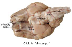
The sheep brain is quite similar to the human brain except for proportion. The sheep has a smaller cerebrum. Also, the sheep brain is oriented anterior to posterior (more horizontally), while the human brain is oriented superior to interior (more vertically.) Materials. Dissection tools and tray, lab gloves, preserved sheep brain. The primary function of the meninges and of the cerebrospinal fluid is to protect the central nervous system. Dura Mater-encases the prain and is the first layer of the brain. Gyrus - a ridge or fold between two clefts on the cerebral surface in the brain. Sulcus - a groove or furrow, especially one on the surface of the brain. Sheep Brain Dissection Key. Image of the brain showing its major features for students to practice labeling. Answers are included. Pretty good picture of the sheep brain labeled. Shows pictures of a sheep and a human brain. Each of the 12 cranial nerves is represented, students color and number each nerve in both brains. October 6, 2012 - Unfortunately, due to the COVID-19 pandemic, the 2021 Brain Bee at MSU will not be held. We look forward to seeing all of our high school students in 2022 · Brain Bee at MSU The 2021 competition will not be held
Black Rabbit, Byker Farm, Ouseburn Valley, Newcastle Upon Tyne, Tyne & Wear, England.
Sheep Brain Dissection Labeled Diagram. angelo. October 16, 2021. Image Result For Sheep Brain Labeled Brain Diagram Human Brain Diagram Brain Anatomy. Sheep Brain External View Labeled Anatomia Veterinaria Anatomia Veterinaria. Pin By Aleena Hanson On A P Brain Anatomy Anatomy Brain Images. Mit Hhmi Summer Workshop For Teachers Neuroanatomy ...
Sheep Brain Dissection Guide - YouTube
The sheep brain is exposed and each of the structures are labeled and described in a sequential manner, in the same way that a real dissection would occur. Sheep Brain Dissection. 1. The sheep brain is enclosed in a tough outer covering called the dura mater. You can still see some structures on the brain before you remove the dura mater.

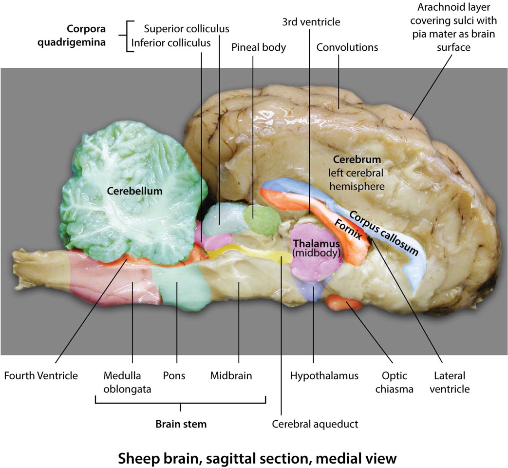
SHEEP BRAIN SAGITTAL VIEW - Biology 220 with Seitchik at ...
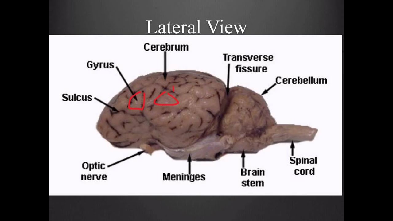
Diagram of Sheep Brain - Lateral view

Practical 3 - Biology 341 with Vaughn at University of ...
Sheep Brain Neuroanatomy Online Self-Test. Use each diagram as a reference, and selected the correct answer for each lettered structure. You may find it useful to open the diagrams in a separate window to review while answering each question. Dorsal Surface.
sheep brain anatomy - YouTube
Sheep Brain Anatomy Lab Manual. Based on original material by R. N. Leaton, Dartmouth College. Contributors to this version: Al Sorenson, Lisa Raskin, Sarah Turgeon, Steve George, and JP Baird. I. Introduction. The brain of the sheep is useful for study because its anatomy is similar to human brain anatomy. Although exact proportions (and names ...
Sheep Brain Dissection Bi - BIOLOGY JUNCTION
About Press Copyright Contact us Creators Advertise Developers Terms Privacy Policy & Safety How YouTube works Test new features Press Copyright Contact us Creators ...

Sheep Brain Dissection Lab
5 3 11 6 22 16 18 1. Gray Matter 2. White Matter 3. Corpus Callosum 4. Lateral Ventricle 5. Caudate Nucleus 6. Septum Pellucidum 7. Fornix 8.

Sheep Brain Dissection Bi - BIOLOGY JUNCTION
Quizlet makes simple learning tools that let you study anything. Start learning today with flashcards, games and learning tools — all for free.

Dorsal View of Sheep Brain
anatomy of the sheep brain video for anatomy class - practice for the practical exam

34 Label The Sheep Brain - Labels Design Ideas 2020
Sheep Brain Dissection The purpose of this exercise is to introduce you to the mammalian brain; you will use a sheep's brain. While the sheep brain differs from the human brain in many details, they both have the same basic anatomy, and, it is larger than the rat brain. Work in teams of four students.

Sheep brain sagittal section medial view - www.anatomynote ...
Start studying Sheep Brain Dissection labeled. Learn vocabulary, terms, and more with flashcards, games, and other study tools.

Pretty good picture of the sheep brain labeled. | Anatomy ...
To study the structure and function of the mammalian (sheep) brain. References: "Nelson Biology 12" (chapter 9), dissection charts, models, internet, other texts. Instructions - External Anatomy: Obtain a handout with diagrams of the brain from your teacher. Working in groups of two or three, select a sheep brain.

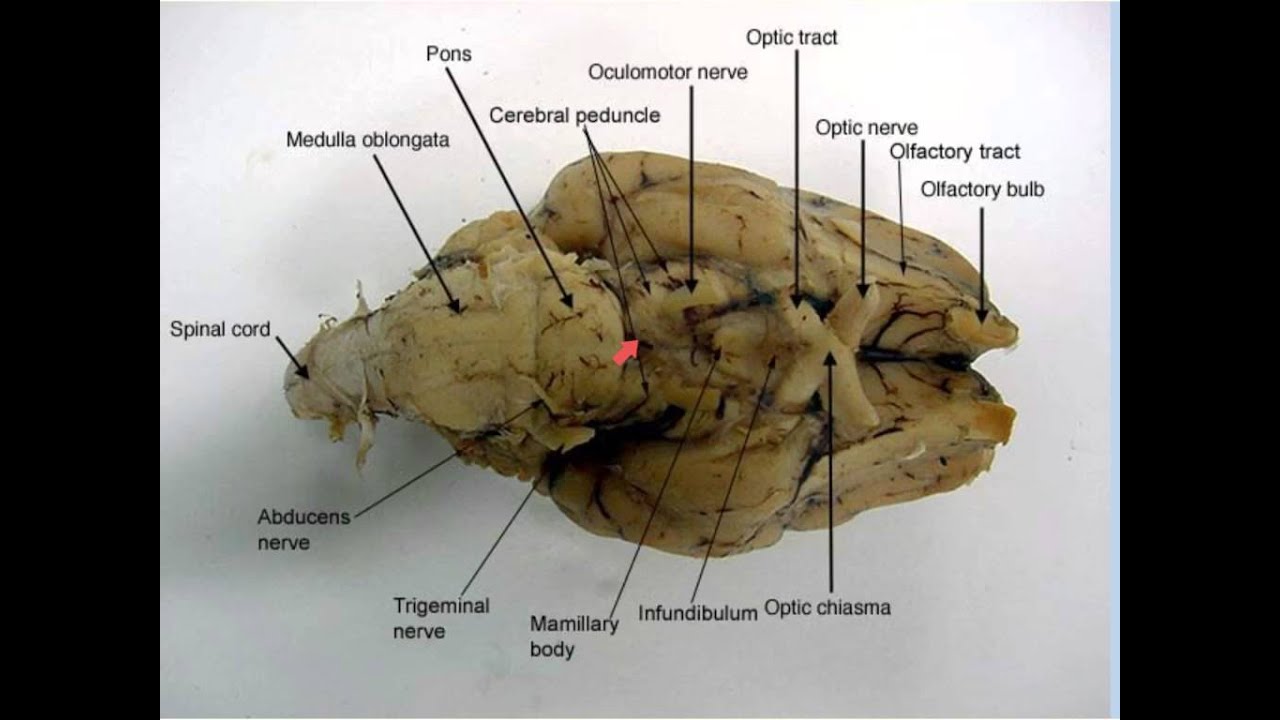
inferior view of sheep brain
Diencephalon and brain stem structures picture. #medical #anatomy #doctor #art #medicine #drawing #health #sketch #nurse #artist #healthcare #illustration ...

Sheep Brain Images
{Label Gallery} Get some ideas to make labels for bottles, jars, packages, products, boxes or classroom activities for free. An easy and convenient way to make label is to generate some ideas first. You should make a label that represents your brand and creativity, at the same time you shouldn't forget the main purpose of the label.

Labeled Diagram Of Sheep Skin | MedicineBTG.com
Sheep Brain. Images taken from the dissection of the sheep's brain, some structures have been labeled. Anatomy Corner resource site for teachers and students of Anatomy and Physiology. Find quizzes, diagrams, and slide presentations on structures, functions, and systems.
Lab Objectives, BIO 2310, Spring 2018 | Clare Hays Biology ...
Dec 14, 2014 - The sheep brain is exposed and each of the structures are labeled and described in a sequential manner, in the same way that a real dissection would occur.

Sheep Brain external view labeled | Brain anatomy ...
Jun 6, 2018 - A virtual sheep brain dissection guides anatomy studies with photos & blank diagrams. Also shop complete dissection kits: guide, ...

sheep brain labeled 6954168 orig - Made By Creative Label
Below are the links to COVID testing information in the county and the state · DOH-Okaloosa site: http://okaloosa.floridahealth.gov/programs-and-services/infectious-disease-services/COVID-19Testing.html

34 Label The Sheep Brain - Labels Design Ideas 2020
sheep brain labeled sheep brain sup label.jpg {Label Gallery} Get some ideas to make labels for bottles, jars, packages, products, boxes or classroom activities for free. An easy and convenient way to make label is to generate some ideas first.

Lab 14 Sheepbraindiss 2
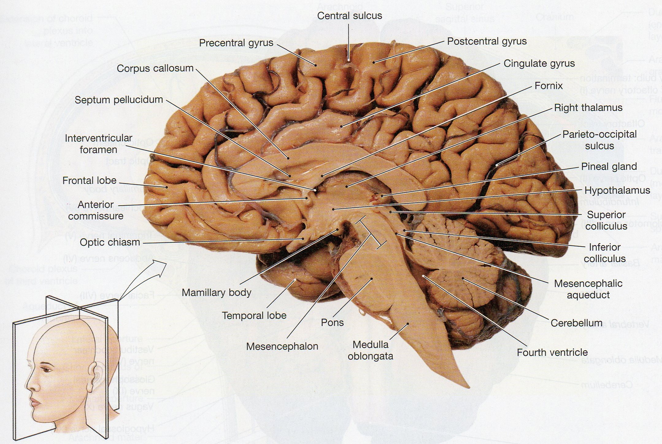
BI 335 - Advanced Human Anatomy and Physiology Western Oregon University Figure 4: Mid-sagittal section of brain showing diencephalon (includes corpus callosum, fornix, and anterior commissure) Marieb & Hoehn (Human Anatomy and Physiology, 9th ed.) - Figure 12.10 Exercise 2: Utilize the model of the human brain to locate the following structures / landmarks for the

Sheep Brain Dissection at Great Bend High School - StudyBlue
labeled brain Ear Anatomy, Brain Anatomy, Human Body Anatomy, ... Sagital Cut Sheep Brain Labeled | sheep brain map midsaggital view labeled diagram of ...

A Skyview Brain-Storm! - NW NOGGIN: Neuroscience outreach ...
Learn about KPU’s plans for January 2022 [Read more] · Spring 2022 Term Update

Labeled Diagram Of Sheep Skin | MedicineBTG.com
San Diego Mesa College offers various certificates and degrees to prepare students for careers relating to Biology.

Sheep Brain at Hillsdale College - StudyBlue
September 15, 2021 - The image below shows a cleanly ... and labeled. ... 14. Finally, a section of the brain is cut to examine the difference between white matter and gray matter. Figure \(\PageIndex{14}\): Cut to show the difference between white and grey matter ... 15. Once you have made the cut like in the above diagram, you should ...

Sheep Brain Dissection Project Guide | Brain anatomy ...
Blood vessels should be dark while all of the myelin coated nerves will be white. Take your time, use the diagrams below, move around gently and you should be able to find most if not all of the cranial nerves. ... . Observe one half of the brain and compare it to the labeled figure below.

Sheep Brain - HUMAN ANATOMY WEB SITE
Play this game to review Human Anatomy. Name this part of the brain. Preview this quiz on Quizizz. Name this part of the brain. Sheep Brain Dissection DRAFT. 6th - 12th grade. 193 times. Biology, Other Sciences. 77% average accuracy. a year ago. mrsturmscience. 0. Save. Edit.

Sheep Brain Diagram Lobes - Data Diagram Medis
Sheep Brain Labeled Diagram. angelo. August 10, 2021. Labeled Brain Brain Anatomy Anatomy And Physiology Anatomy. Image Result For Sheep Brain Labeled Nursing Study Guide Anatomy And Physiology Nursing Study In 2021 Nursing Study Guide Nursing Study Human Brain Anatomy.

Diagram of Sheep Brain - Inferior view
function, and pathology. Those students participating in Sheep Brain Dissections will have the opportunity to dissect and compare anatomical structures. At the end of this document, you will find anatomical diagrams, vocabulary review, and pre/post tests for your students. The following topics will be covered: 1.

Dissecting Sheep Brains With Sixth Graders | Brains Explained
Labeled Diagrams of the Human Brain. Central Core. The central core consists of the thalamus, pons, cerebellum, reticular formation and medulla. These five regions are the central areas that regulate breathing, pulse, arousal, balance, sleep and early stages of processing sensory information. The thalamus interprets the sensory information and ...
Sheep Brain Dissection Lab
Picture Nerve Anatomy, Brain Anatomy, Human Anatomy And Physiology, Medical Anatomy, Neuron ... Sheep Brain Dissection Project Guide | HST Learning Center.

Sheep brain exterior | Brain science, Life science middle ...
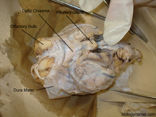
Label theBrain of the Sheep. Publisher: Biologycorner.com; follow on Google+ This work is licensed under a Creative Commons Attribution-NonCommercial 3.0 Unported License. Brain Label Answer Key. Image adapted from a photograph of the sheep brain. ...
20 New Lateral View Of Sheep Brain Labeled
Labeled Sheep Heart Picture #1. Explore nicellian's photos on Flickr. nicellian has uploaded 157 photos to Flickr.

Labeled Diagram Of Sheep Skin | MedicineBTG.com
This is an online quiz called Sheep Brain. This quiz has tags. Click on the tags below to find other quizzes on the same subject. Anatomy. sheep brain.

Pin on Work: Nervous System
DISSECTION OF THE SHEEP'S BRAIN Introduction The purpose of the sheep brain dissection is to familiarize you with the three-dimensional structure of the brain and teach you one of the great methods of studying the brain: looking at its structure. One of the great truths of studying biology is the saying that "anatomy precedes physiology".

sheep brain | Dorsal View of Sheep Brain | Brain anatomy ...

Brain Flashcards | Quizlet

Sheep Brain Dissection Lab Sheet | Brain diagram, Brain ...

Sheep Brain Dissection Guide with Pictures | Secondary ...

Brain - HUMAN ANATOMY WEB SITE

Sheep Brain Diagram Unlabeled | World of Diagrams

Sheep Brain
Sheep Brain Dissection Project Guide | HST Learning Center
Physiological Psychology
0 Response to "43 sheep brain labeled diagram"
Post a Comment