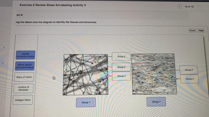
40 drag the labels onto the diagram to identify the types of connective tissue proper.
Connective Tissue - Definition, Types, and Functions As the name suggest connective tissue is a tissue that connects the different cell and structure of the body. Also, these tissues perform other function that helps in the various mechanism of the body. Drag the labels onto the diagram to identify the cell types and matrix components of areolar connective tissue, a model connective tissue. Of the two major cell types found in nervous tissue, __________ cells are highly specialized to generate and conduct electrical signals.
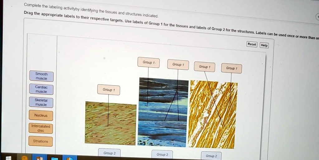
These tissues include the skeletal muscle fibers blood vessels nerve fibers and connective tissue. Drag the labels onto the diagram to identify structural features associated with skeletal muscle. Drag the labels onto the diagram to identify the blood types that correspond to specific blood typing test results.

Drag the labels onto the diagram to identify the types of connective tissue proper.
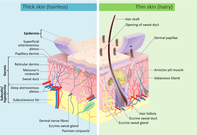
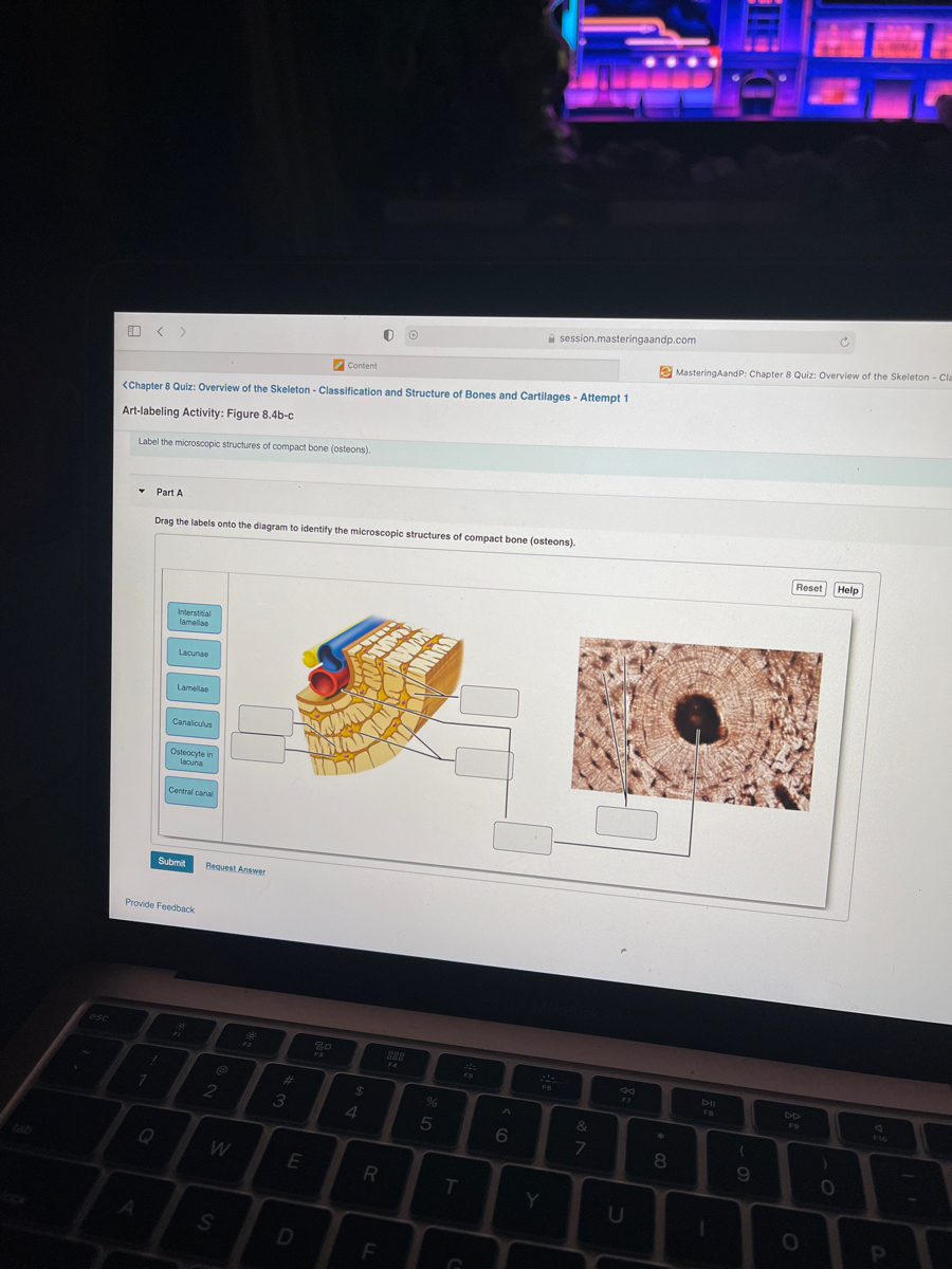
Question: Drag the labels onto the diagram to identify the components of the integumentary system. Reset Stre Cutaneous embra Har shaft Pore swear gland duct Talle COCOLISCIA Seondecus gland. rectomp muscle Hair follicle cam QUISA Swearglana du Sweat gland Nerve fibers Submit Previous Answers Request Answer . There are six types of connective tissue found in the human body: Loose Connective Tissue- as its name suggests, the cells of this tissue are scattered with loose fibers in its matrix.It lies under the skin and in between organs. the main function of loose connective tissue is to provide nutrition and prevent a shock or injury to the nearby organs, to fight against infection, hold organs ... Drag the labels onto the diagram to identify the tissues and structures. Reset Help central cand matrix Group 2 lacuna Group 2 Group 2 osteocyte in lacuna Group 2 C chondrocyto Group 2 bono (osseous tissue) Group 1 Group 1 hyaline cartilago 2 See answers Advertisement Advertisement mistinat mistinat Answer: See image. Explanation: Advertisement Advertisement marianaegarciaperedo ...
Drag the labels onto the diagram to identify the types of connective tissue proper.. Image: Drag the labels onto the diagram to identify the types of connective tissue proper. Drag the correct description under each cell structure to identify the role it plays in the cell. Part a animal cell structure drag the labels onto the diagram to identify the structures of an animal cell. Drag the labels onto the diagram to identify the parts of the cell. Labels can be used once more than once or not at all. Connective tissue Basement membrane Nucleus IDIO Lumen of duct S tified cuboidal cells Submit Request Answer 1301051aafd2 787. This problem has been solved! See ... The structure of bone tissue suits the function. Drag the labels onto the diagram to identify structural features associated with skeletal muscle. Study questions on anatomy review. Drag the appropriate labels to their respective targets. Drag the labels onto the diagram to identify the blood types that correspond to specific blood typing test ...
Drag the labels onto the diagram to identify the types of connective tissue proper. look at pic When a molecule is _______ it loses electrons, and when a molecule is ______ it gains electrons. Connective tissue is a term used to describe the tissue of mesodermal origin that that forms a matrix beneath the epithelial layer and is a connecting or supporting framework for most of the organs of the body. This lab will focus on the so-called connective tissue proper and cartilage; the next lab will focus on bone. Drag the labels onto the diagram to identify the types of connective tissue proper. areolar tissue, adipose tissue, reticular tissue, dense regular connective tissue, dense irregular connective tissue, elastic tissue. Myosatellite cells are found in association with _____ muscle. skeletal . What is the only type of cell found in cartilage matrix? (Figure 4-16) chondrocytes. The function of ... The four basic types of tissue are epithelial tissue, connective tissue, muscle tissue, and nervous tissue. If a single, broad functional term were assigned to each basic tissue, the terms would be covering (epithelial tissue), support (con-nective), movement (muscle), and control (nervous). However, these terms reflect only a fraction of the functions that each tissue performs. Chapter 4 ...
Name the major types of fibers and molecules found in the extracellular matrix of the skeletal system. Collagen ... thinner connective tissue membrane that lines the surface of the medullary cavity. 4. Describe the structure of compact bone. ... All bone growth occurs by the apposition of new bone lamellae onto existing bone or other CT. As ... Chapter 4 Notes Epithelial and Connective Tissue. Histology: the study of tissues. 1. Definition. a tissue is a group of similar cells with intercellular material that together perform a specific function. Identify the type and tissue shown. Loose Connective Tissue. Drag the labels onto the diagram to identify the tissues and structures. Identify the structure at the tip of the pointer. Nucleus. Identify the ground substance for this tissue. Plasma. The epithelial tissue shown attaches to a _________ membrane. Basement. Question: Drag the labels onto the diagram to identify structural features of epi the lium. Connective tissue Basement membrane Nucleus IDIO Lumen of duct S ... Transcribed image text: Art-labeling Activity: Classifying Epithelia Drag the labels onto the diagram to identify the types of epithelia. Color breathing is a simple stress reducing activity that may be quickly learned.
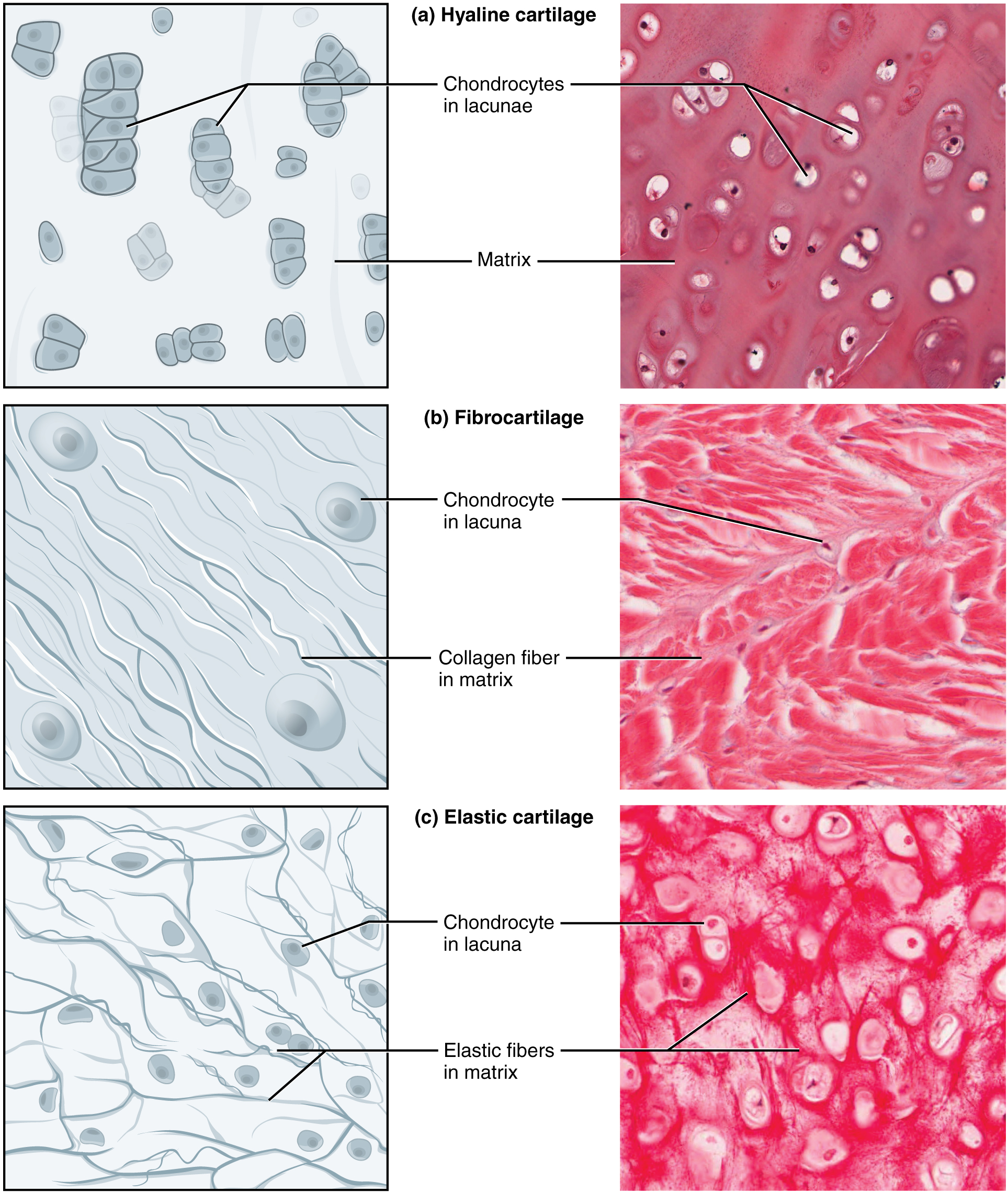
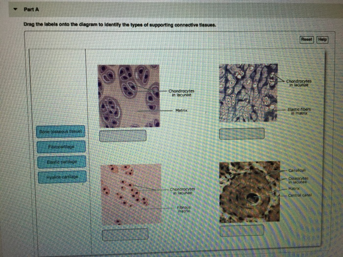
Part A Drag the labels onto the diagram to identify the types of supporting connective tissues. ANSWER: Correct Chapter 4 Book-specific Clinical Case Activity All her life Anne Marie has felt "delicate." She is a horrible athlete. She once dislocated both shoulders attempting to play high school volleyball. And she has an unstable feeling, as if her kneecaps will dislocate when she tries ...

Identify the connective tissue proper cellular component labeled "F". ... Drag the labels onto the diagram to identify the muscle types based on fascicle ...

For page 1, the answers are:- Aerolar tissue Adi …. View the full answer. Transcribed image text: -Aibrocytes Reticular fbers bundles Part A Drag the labels onto the diagram to identify the types of supporting connective tissues Chondrocytes in lacunae Chondrocytes in lacunae Matrix Elastic fibers Böne Osteocytes in lacuna Chondrocytes in ...
Drag the labels onto the diagram to identify the types of supporting connective tissues. look at pic A marked loss in strength and elasticity of connective tissue characterizes Marfan's syndrome.
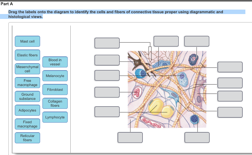
Drag the labels onto the diagram to identify the cells and fibers of connective tissue proper using diagrammatic and histological views. Image: Reticular Fibers ...

View Homework Help - chapter 4.pdf from AAS a&p 198 at University of New Hampshire. 12/24/13 chapter 4 chapter 4 Due: 9:00am on Thursday, September 26, 2013 You will receive no credit for items you

Drag the labels onto the diagram to identify the types of connective tissue proper. (week three assignment one) #7. Drag the labels onto the four types of tissue membranes. ... Drag the labels onto the diagram to identify the effects of isotonic, hypotonic, and hypertonic solutions on red blood cells.

Identify the tissue type and a location where it is found. Loose Areolar Connective Tissue •Papillary layer of dermis • Hypodermis •Around organs • Basement membrane of mucous membranes •Surrounding blood vessels Blood Vessel. Identify the tissue type and its function.

Drag the labels onto the diagram to identify the cells and fibers of connective tissue proper using diagrammatic and histological views. This problem has been ...
And muscle tendons are made of dense regular connective tissue, though in ligaments not. / physical therapy in perrysburg for . These consist of the arm, located between the shoulder and elbow joints; Ap1 Lab 9 10 Hw Flashcards Quizlet from quizlet.com Drag the labels onto the diagram to identify the various types of synarthroses and. The ligaments, joint capsules and labrum are fixed ...
100% (5 ratings) Answer There types of tissues included in connective tissue proper are : Loose (areolar) con …. View the full answer. Transcribed image text: Drag the labels onto the diagram to identify the cells and fibers of connective tissue proper using diagrammatic and histological views. Previous question Next question.
Meiosis 2 of 3. Drag the labels onto the diagram to identify the stages in which the lagging strand is synthesized. Chapter 7 The Skeletal System Bone Tissue Tour of an animal cell organelle functions can you identify the functions of the parts of an animal cell. Drag the labels onto the diagram to identify the parts of the cell. After each ...

See Page 1. skeletal cardiac smooth. Label the cell types and matrix components of areolar connective tissue, a model connective tissue. Help Reset Squamous cell Simple Cuboidal cell Apical surface Stratified Basal surface Columnar cell. 2/18/2020 Histology - Lab 4/12 Part A Drag the labels onto the diagram to identify the cell types and matrix ...
Transcribed image text: Part A Drag the labels onto the diagram to identify the cells and fibers of connective tissue proper using diagrammatic and histological views. Reset Help Mast cell Blood in vesse Free macrophage Melanocyte Lymphocyte IIIII Fibroblast Ground substance 108 III Elastic fibers Adipocytes Collagen fibers Reticular fibers Mesenchymal 001 Fixed macrophage Submit Request Answer

The major types of connective tissue are connective tissue proper, supportive tissue, and fluid tissue. Loose connective tissue proper includes adipose tissue, areolar tissue, and reticular tissue. These serve to hold organs and other tissues in place and, in the case of adipose tissue, isolate and store energy reserves.

(c) Connective tissue proper: loose connective tissue, reticular Description: Network of reticular fibers in a typical loose ground substance; reticular cells lie on the network. Function: Fibers form a soft internal skeleton (stroma) that supports other cell types including white blood cells, mast cells, and macrophages.

Part A Drag the labels onto the diagram to identify the types of epithelia. ANSWER: Correct Art-labeling Activity: Types of Connective Tissue Proper Learning Goal: To learn the types of connective tissue proper. Label the types of connective tissue proper.
Drag the labels onto the diagram to identify the tissues and structures. Reset Help central cand matrix Group 2 lacuna Group 2 Group 2 osteocyte in lacuna Group 2 C chondrocyto Group 2 bono (osseous tissue) Group 1 Group 1 hyaline cartilago 2 See answers Advertisement Advertisement mistinat mistinat Answer: See image. Explanation: Advertisement Advertisement marianaegarciaperedo ...
There are six types of connective tissue found in the human body: Loose Connective Tissue- as its name suggests, the cells of this tissue are scattered with loose fibers in its matrix.It lies under the skin and in between organs. the main function of loose connective tissue is to provide nutrition and prevent a shock or injury to the nearby organs, to fight against infection, hold organs ...
Question: Drag the labels onto the diagram to identify the components of the integumentary system. Reset Stre Cutaneous embra Har shaft Pore swear gland duct Talle COCOLISCIA Seondecus gland. rectomp muscle Hair follicle cam QUISA Swearglana du Sweat gland Nerve fibers Submit Previous Answers Request Answer .


0 Response to "40 drag the labels onto the diagram to identify the types of connective tissue proper."
Post a Comment