45 label the brain anatomy diagram answers
10-06-2021 · Label a diagram if you're a visual learner. If you learn best by reading and re-reading, or by looking at diagrams, use print-outs of the muscular system to help you study. Read over the labeled diagram carefully, then switch to a blank copy of the same diagram and try to fill in the names of as many muscles as you can remember. Brain 101 4. Pre-Quiz - Part 2. For each statement, decide whether it is a function of the: 1. Brain Stem 2. Cerebellum . 3. Occipital Lobes 4.
WebMD's Brain Anatomy Page provides a detailed diagram and definition of the brain including its function, parts, and conditions that affect it.

Label the brain anatomy diagram answers
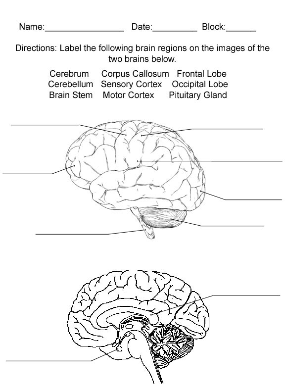
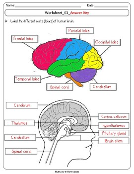
Answers: Label the Brain Diagram The Brain Read the definitions below, then label the brain anatomy diagram. Cerebellum - the part of the brain below the back of the cerebrum. It regulates balance, posture, movement, and muscle coordination. Corpus Callosum - a large bundle of nerve fibers that connect the left and right cerebral hemispheres. Answers: Neuron Anatomy: Label the Cell Label the axon, dendrites, cell body, nucleus, Schwann's cells, and nodes of Ranvier. Answers: Skin Anatomy See the layers of the skin and the glands (and other tissues) it contains. Skin Anatomy: Label Me! Printout Label the skin anatomy diagram. Answers: Spine and Skull Anatomy Diagram Label the spine ... Browse Printable 5th Grade Science Worksheets. Award winning educational materials designed to help kids succeed. Start for free now!
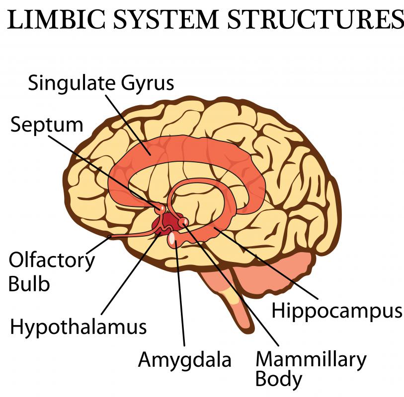
Label the brain anatomy diagram answers. 2,687 labeled brain anatomy stock photos, vectors, and illustrations are available royalty-free. See labeled brain anatomy stock video clips. of 27. brain diagram with labels hypothalamus vector brain diagram pons cerebrum and cerebellum brain pons brain anatomy amygdala brain labelled amygdala brain human midbrain diagram pons. BRAIN ANATOMY FUNCTION CHEAT SHEET System or Part Function Misc. Brainstem Responsible for automatic survival reflexes Spinal Cord Controls simple reflexes Pathway to neural fibers Medulla Controls/regulates heartbeat and breathing To and from brain Reticular Formation Helps control arousal, responds to change in monotony ... Mid Sagittal Brain Unlabeled Hand Crochet Decorative Plates Decor. Beware Of Amygdala Hijacks Brain Diagram Human Brain Parts Brain Parts. Shame Cube In 2021 Brain Diagram Human Brain Diagram Human Brain. The Wired Mind Virginia Magazine Human Brain Diagram Brain Diagram Basic Anatomy And Physiology. Labeled Pictures Of The Brain Labeled Brain ... Continue with more related things such sheep brain diagram labeled, labeled sheep brain worksheet and label the brain anatomy diagram answers. We hope these Brain Diagram Worksheet photos gallery can be a direction for you, bring you more ideas and most important: help you get what you need.
Label the organ systems underneath each illustration and label the selected organs by using the terms available. When you finish, select different colors for each organ system andcolor them in. ORGAN SYSTEMS (CONTINUED) The skinand other structures are in the integumentarysystemand the digestive systeminvolves the breakdown and absorption ... The cerebellum ("little brain") is a fist-sized portion of the brain located at the back of the head, below the temporal and occipital lobes and above the brainstem. Like the cerebral cortex, it has two hemispheres. The outer portion contains neurons, and the inner area communicates with the cerebral cortex. May 15, 2011 · What Cortical Region of the brain would these doctors be stimulating? 20. Let’s practice!!! Read the definitions, then label the brain anatomy diagram. 21. Answers 22. Read the definitions, then label the neuron diagram below. 24. Talking related with Brain Label Worksheet, below we will see particular related photos to inform you more. human brain diagram blank, label the brain anatomy diagram answers and plant cell diagram with labels are three of main things we want to show you based on the post title.
Label The Brain Anatomy Diagram Quiz. angelo on November 23, 2021. Activity The Brain The Brain For Kids Brain Activities Human Body Activities. Mri Sagittal Anatomy Of Brain Level 1 Mri Is Sensitive To Changes In Cartilage And Bone Structure Resulting From Inju Brain Anatomy Mri Brain Radiology Student. Pin By Line Brandt On Health Brian ... Respiratory system lesson for kids. The lesson shows animation of ventilation and respiration. It covers the journey of air into the lungs, and the transfer of oxygen from the alveoli sacs to the red blood cells in the capillaries. Transcribed image text: BRAIN WORKSHEET Read the definitions below, then label the brain anatomy diagram Lateral View of the Brain EnchantedLearning.com Cerebellum - the part of the brain below the back of the cerebrum. It regulates Parietal Lobe of the Cerebrum - the middle balance, posture, movement, and muscle lobe of each cerebral hemisphere between the coordination. Benchmark: SC.912.L.14.26 AA Identify the major parts of the brain on diagrams or models. Benchmark Clarification: Students will identify the major parts of the brain on diagrams. Content Limit: Items are limited to the cerebrum, cerebellum, pons, medulla oblongata, brain stem, frontal lobe, parietal lobe, occipital lobe, and temporal lobe.
Labeled Diagrams of the Human Brain Central Core The central core consists of the thalamus, pons, cerebellum, reticular formation and medulla. These five regions are the central areas that regulate breathing, pulse, arousal, balance, sleep and early stages of processing sensory information.
WebMD's Heart Anatomy Page provides a detailed image of the heart and provides information on heart conditions, tests, and treatments.
Start studying exam 4 anatomy and physiology. Learn vocabulary, terms, and more with flashcards, games, and other study tools.
Import a diagram: Add a new diagram tab as described above, and simply drag the diagram file from your device onto the canvas to import it. 1/5/2019 · Drag the labels on the left onto the diagram to identify the compounds that couple each stage. 13/4/2017 · Drag the label onto the diagram to identify the stages of cellular respiration 2 See ...
Start studying Label the Brain Anatomy Diagram. Learn vocabulary, terms, and more with flashcards, games, and other study tools.
The Human Brain Coloring Book Awesome 11 Best Of Cadaver Brain Label Worksheet Brain Brain Diagram Brain Anatomy Coloring Pages . Brain Labeling Brain Challenge Parts Of The Brain Labels . Shame Cube In 2021 Human Brain Diagram Brain Diagram Human Brain . Human Brain Worksheets Superstar Worksheets Human Brain Human Body Videos Biology ...
Diagram Worksheets. Label the Parts of a Sheep Brain. Print out these diagrams and fill in the labels to test your knowledge of sheep brain anatomy. Internal anatomy: label the right side (.pdf) External anatomy: label the top view (.pdf) External anatomy: label the bottom view (.pdf) What other users say: Fun and Educational.
Try our top 10 quizzes : 1 - the skeleton: test your knowledge of the bones of the full skeleton. 2 - the brain: can you name the main anatomical areas of the brain?. 3 - the cell: learn the anatomy of a typical human cell. 4 - the skull: Do you know the bones of the skull?. 5 - the axial skeleton: How about the bones of the axial skeleton?. 6 - the heart: name the parts of the human heart
Think back to last Halloween for a minute. Wherever you looked, there were vampires, ghosts, or bony skeletons grinning back at you. Vampires and ghosts don't really exist, but skeletons sure do! Every single person has a skeleton made up of many bones. These bones give your body structure, let you ...
05-06-2014 · Defining circuit function requires knowledge of circuit structure. Three levels of anatomy should be considered: long-range, intermediate-range, and detailed connectivity. 1b-i. Long-Range Connectivity. Traditional neuroanatomy has focused on large-scale, long-range connections between different brain regions (e.g. the thalamocortical tract).
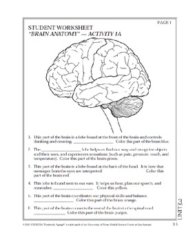
This brain part controls thinking. This brain part controls balance, movement, and coordination. This brain part controls involuntary actions such as breathing, heartbeats, and digestion. This part of the nervous system moves messages between the brain and the body. This part of the cerebrum interprets and sorts information from the senses.
BI 335 - Advanced Human Anatomy and Physiology Western Oregon University Figure 4: Mid-sagittal section of brain showing diencephalon (includes corpus callosum, fornix, and anterior commissure) Marieb & Hoehn (Human Anatomy and Physiology, 9th ed.) - Figure 12.10 Exercise 2: Utilize the model of the human brain to locate the following structures / landmarks for the
Image of the brain showing its major features for students to practice labeling. Answers are included.
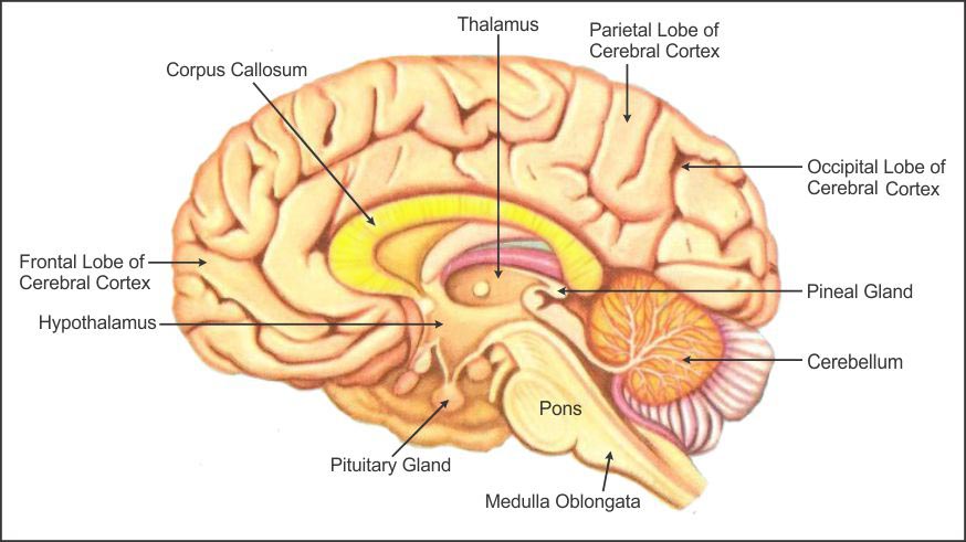
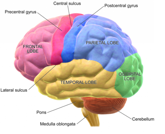
Labeled brain diagram. First up, have a look at the labeled brain structures on the image below. Try to memorize the name and location of each structure, then proceed to test yourself with the blank brain diagram provided below. Labeled diagram showing the main parts of the brain.
Brain Quiz. Choose a viewpoint from the drop-down menu. Description. Scores: View Total; Correct / Attempts: 0 / 0: 0 / 0: Explore the brain inside and out! Identify the parts of the brain and learn their functions. Can you get them all right? Programmed by Quinn Baetz. Part of the curriculum unit: ...
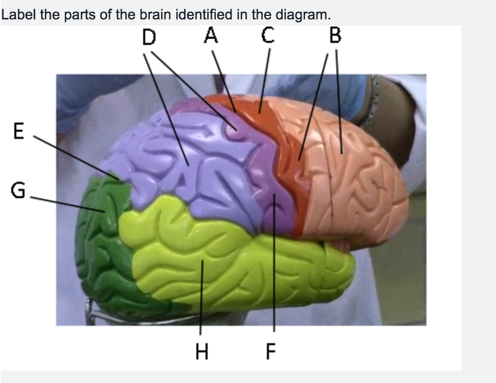
Label the Brain Anatomy Diagram. The Brain. Read the definitions below, then label the brain anatomy diagram. Cerebellum - the part of the brain below the back of the cerebrum. It regulates balance, posture, movement, and muscle coordination. Corpus Callosum - a large bundle of nerve fibers that connect the left and right cerebral hemispheres.
15-11-2018 · Write your answers in boxes 32-38 on your answer sheet. How the elephants sense these sound vibrations is still unknown, but O’Connell-Rodwell, a postdoctoral researcher at Stanford University, proposes that elephants are ‘listening’ with their 32 by two kinds of nerve endings that respond to vibrations with both 33 frequency and slightly higher frequencies.
Label The Skin Anatomy Diagram Answers 1/4 Read Online Label The Skin Anatomy Diagram Answers streaming.missioncollege.org 4, Label the skin structures and areas indicated in the accompanying diagram of skin. Hair shaft 006 /DÉRv)11s Subcutaneous ssue, or PACINlHî (deep pressure receptor) 50 6, What substance is manufactured in the skin (but ...
This is an online quiz called Label Parts of the Brain. There is a printable worksheet available for download here so you can take the quiz with pen and paper. From the quiz author. Okay. This quiz has tags. Click on the tags below to find other quizzes on the same subject. biology. brain. Your Skills & Rank. Total Points. 0.
Label The Parts Of The Brain Worksheet Worksheets For All Brain Parts Brain Diagram Brain. Brain Structure And Function Worksheet Worksheets Are A Crucial Portion Of Researching English Infant In 2021 Brain Diagram Brain Anatomy Brain Anatomy And Function. Brain Parts Fill In The Blank Color Nervous System Lesson Biology Worksheet Teaching ...
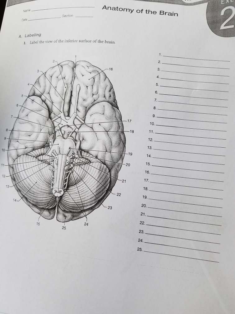
Study guide for the digestive system focusing on vocabulary and labeling diagrams; intended for high school students taking anatomy and physiology. Shows pictures of a sheep and a human brain. Each of the 12 cranial nerves is represented, students color and number each nerve in both brains.
An increase in ____ in the blood − and a resulting drop in pH − signals the breathing control center in the brain to increase the breathing rate. This keeps gas exchange in tune with body needs. The right side of the heart pumps blood returning from body tissues to ________ in the lungs.
The activity includes an external view of the brain where students label the lobes of the cerebrum (frontal, parietal, occipital, and temporal) and the cerebellum. Next students drag and drop labels to the internal structures, such as the thalamus, midbrain, corpus callosum, pineal body, and colliculi.
Browse Printable 5th Grade Science Worksheets. Award winning educational materials designed to help kids succeed. Start for free now!
Answers: Neuron Anatomy: Label the Cell Label the axon, dendrites, cell body, nucleus, Schwann's cells, and nodes of Ranvier. Answers: Skin Anatomy See the layers of the skin and the glands (and other tissues) it contains. Skin Anatomy: Label Me! Printout Label the skin anatomy diagram. Answers: Spine and Skull Anatomy Diagram Label the spine ...
Answers: Label the Brain Diagram The Brain Read the definitions below, then label the brain anatomy diagram. Cerebellum - the part of the brain below the back of the cerebrum. It regulates balance, posture, movement, and muscle coordination. Corpus Callosum - a large bundle of nerve fibers that connect the left and right cerebral hemispheres.













0 Response to "45 label the brain anatomy diagram answers"
Post a Comment