0 cardiac cycle diagram labeled
the cardiac cycle. Quiz Questions #3a, 4a, 5a, 6a: Cardiac Cycle Phase • These question asks you to view a diagram of the heart and predict what stage of the cell cycle it's in. You may take notes on the diagrams below. The dye-labeled blood has been colored light here to make it more visible. Quiz Question #3b, 4b, 5b, 6b: Cardiac Cycle Graphs Download Diagram of the phases of cardiac cycle with main parts labeled. Circulation of blood through the heart. Vector illustration in flat style over white background. Stock Vector and explore similar vectors at Adobe Stock.
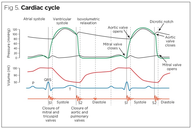
Dec 09, 2016 · The cardiac cycle diagram shown to the right depicts changes in aortic pressure (AP), left ventricular pressure (LVP), left atrial pressure (LAP), left ventricular volume (LV Vol), and heart sounds during a single cycle of cardiac contraction and relaxation. These changes are related in time to the electrocardiogram.

Cardiac cycle diagram labeled
The cardiac cycle is defined as a sequence of alternating contraction and relaxation of the atria and ventricles in order to pump blood throughout the body. It starts at the beginning of one heartbeat and ends at the beginning of another. The process begins as early as the 4th gestational week when the heart first begins contracting.. Each cardiac cycle has a diastolic phase (also called ... The cardiac cycle has 2 main phases, systole and diastole, defined by whether the heart is depolarized and contracting vs repolarized and relaxed. This post will walk you through one cycle of the heart using a step-by-step diagram that will provide you with easy notes and a simple explanation of cardiac physiology. Diagram: Blood flow through the heart, cardiac circulation pathway steps, and cardiac anatomy and structures. Blue arrows (deoxygenated blood); Red arrows (oxygenated blood). Now that we have a good understanding of the blood flow through the heart using the cartoon diagrams, we can apply it to a more realistic image of the heart.
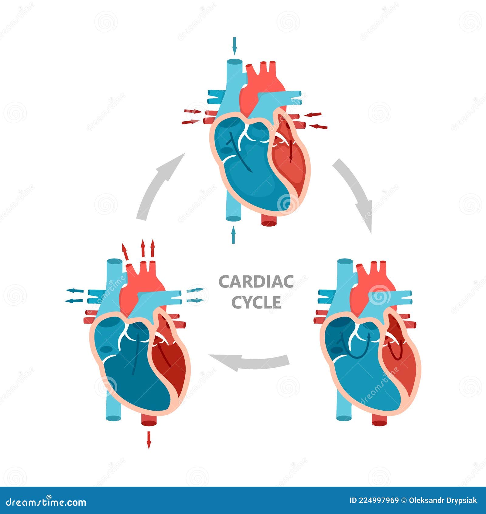
Cardiac cycle diagram labeled. Cardiac cycle of the left side of the heart. The electrocardiogram (EKG) below the diagram shows the corresponding waves with each phase of the cardiac ... (See Wiggers diagram, which presents the stages, label-wise, in 3,4,1,2 order, left-to-right.) ... Label the Wiggins diagram. You should be able to label a figure like this and relate it to the conducEon aspects of the cardiac cycle. Describe what is occurring in each panel of this diagram. Be able to label all the parts of the heart and associated blood vessels. Jan 06, 2022 · The cardiac cycle is the period between the start of one heartbeat and the start of another. One cycle is complete when the heart fills with blood and then ejects the blood. The cardiac cycle has ...
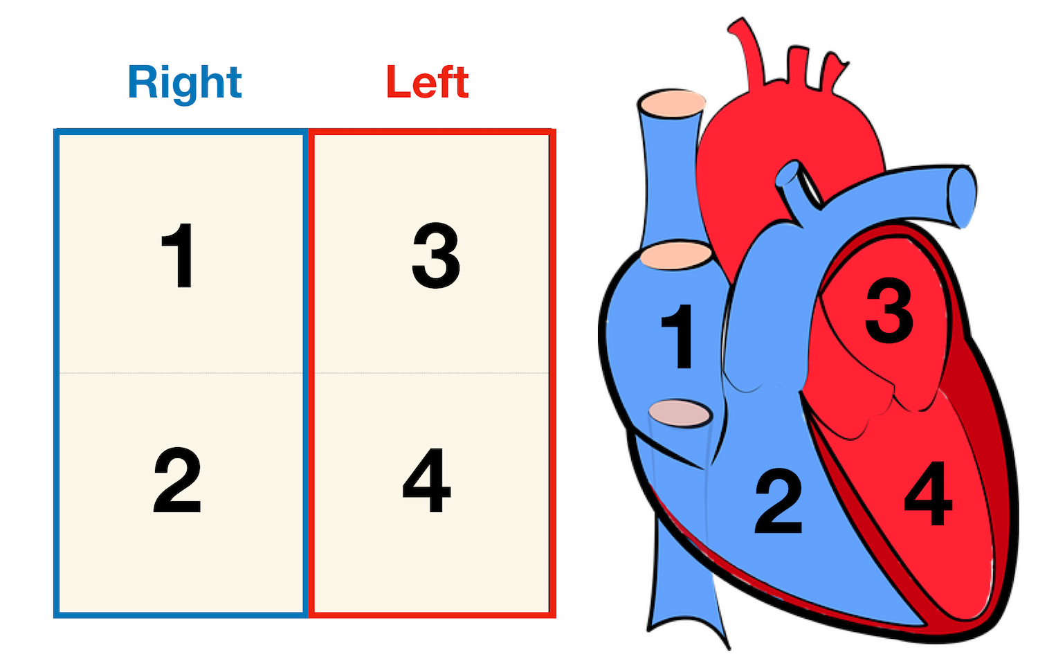
Image: Cardiac anatomy diagram showing the right and left side of the heart. The right side includes chambers 1 and 2. The left side includes chambers 3 and 4. Top vs Bottom of the Heart Next, we can divide the top 2 chambers of the heart from the bottom 2 chambers. The 2 chambers on top are known as the atria, and they include boxes 1 and 3. Cardiac Cycle – Summary and Wigger's Diagram · Opening and closing of valves · Role of atrial contraction in Ventricular filling · Isovolumetric ... The Cardiac Cycle III: Volumes in the Left Ventricle: 6 mins: 0 completed: Learn. The Cardiac Cycle IV: Heart Sounds: 7 mins: 0 completed: Learn. The Cardiac Cycle V: The Wiggers Diagram: 10 mins: 0 completed: Learn. The Cardiac Cycle VI: The Ventricular Pressure-Volume Loop: 9 mins: 0 completed: Learn. Cardiac Output I: Definitions: 5 mins: 0 ... LC: Label the heart and describe the cardiac cycle Starter: 1. Which side of the heart contains oxygenated blood? 2. Which side of the heart has is larger 3. Can you explain why it is larger? 4. What is the purpose of valves?
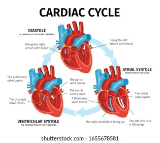
Module 1: Anatomy and Physiology of the Heart Page 10 Developed by Tony Curran (Clinical Nurse Educator) and Gill Sheppard (Clinical Nurse Specialist) Cardiology (October 2011) The four heart valves open and close in response to pressure changes that occur in the ventricles during each cardiac cycle. Anatomy: Images. Cardiac Conduction. Lewis (1918) Gray's Anatomy 20th ed (in public domain at Yahoo or BartleBy) Cardiac Cycle (Wiggers Diagram) Physiology: Nerve Impulse Transmission. Nerve Impulse (Action Potential) See Nerve Impulse; As with Neurons, specialized cardiac Muscle transmits electrical signals The cardiac cycle is a series of contractions that ensure blood is flowing in the correct direction. The cycle is broken down into three stages: • Cardiac Diastole - the entire heart is relaxed • Atrial Systole, also known as Ventricular Diastole • Ventricular Systole, also known as Atrial Diastole . Cardiac Diastole. In cardiac ... View Lab11_heart.pdf from HS 2300 at Western University. Kin 2222A / HS 2300 Lab 12: Vessels, Heart & Cardiac Cycle pumping nutrients, beat by beat OBJECTIVES: By the end of this lab, you should be
2.3.1 Blood circulatory system - Siyavula: Life Sciences ...
cardiac cycle duration increases with a decrease in the heart rate and on the other hand it shortens with increasing heart rate. At a normal heart rate of 75 beats per minute, one cardiac cycle lasts 0.8 second. Under resting conditions, systole occupies ⅓ and diastole ⅔ of the cardiac cycle duration. At an increasing heart rate

4 Phases of the Cardiac Cycle Diagram | Quizlet
The cardiac cycle is the sequence of events that occurs when the heart beats. As the heart beats, it circulates blood through pulmonary and systemic circuits of the body. There are two phases of the cardiac cycle: The diastole phase and the systole phase. In the diastole phase, heart ventricles relax and the heart fills with blood.

The Cardiac Cycle Demystified
THE CARDIAC CYCLE Objectives: Identifying Factors which affect heart rate Describe Cardiac Functional Anatomy (including a review of blood flow and valves) Understand the Wiggers Diagram of Cardiac Cycle Differentiate between Wiggers Diagram and the Pressure Volume Curve Review the electrical basis of excitable cardiac tissue

Wednesday 20th November - COPY: Objective To understand how ...
The period of timethat begins with contraction of the atria and ends with ventricular relaxation is known as the cardiac cycle (Figure 19.3.1).The period of contraction that the heart undergoes while it pumps blood into circulation is called systole.The period of relaxation that occurs as the chambers fill with blood is called diastole.Both the atria and ventricles undergo systole and diastole ...
Pressure-Volume Loop Relationships | Adam Goodwill ...
This page includes the following topics and synonyms: Cardiac Electrophysiology Anatomy, Cardiac Conduction System, Heart Conduction Anatomy, Sinoatrial Node, SA Node, Atrioventricular Node, AV Node, Atrioventricular Bundle, AV Bundle, Bundle of His, Kent-His Bundle, Atrioventricular Fasciculus, Purkinje Fiber, Cardiac Cycle, Wiggers Diagram.

Cardiac Cycle Diagram from Diastole to Systole: Phases of ...
THE CARDIAC CYCLE 1. The cardiac cycle is the sequence of events in one heartbeat. In its simplest form, the cardiac cycle is the simultaneous contraction of both atria, followed a fraction of a second later by the simultaneous contraction of both ventricles. 2. The heart consists of cardiac muscle cells that connect with each other - they are branched - and
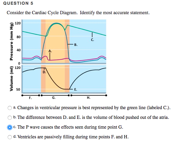
Solved Consider the Cardiac Cycle Diagram. Identify the most ...
This diagram remains the most commonly used model to teach the cardiac cycle today. A typical Wiggers diagram is shown in Fig. 1. Fig. 1. The Wiggers diagram. From top to bottom, the lines show: 1) aortic pressure, 2) ventricular pressure, 3) atrial pressure, 4) electrocardiogram, 5) mitral and aortic valve opening and closing, and 6) heart sounds.
Cardiac Cycle - Summary and Wigger's Diagram | Epomedicine
Cardiac Cycle. I. Structure/Function Relationships. A. Atria. 1. On the diagram below, name & label the fibers associated with the individual atria. 2. On the diagram below, name & label the fibers that wrap around both atria. a) What is the function of both types of atrial fibers? 3.

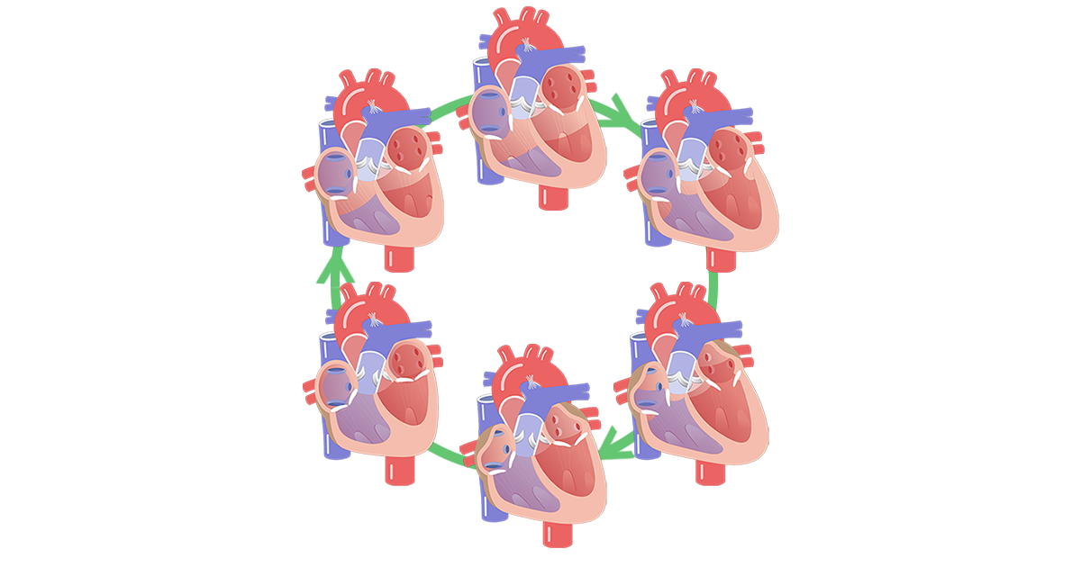
Two cardiac cycles diagram. The color bar illustrates seven ...
Start studying The Cardiac Cycle Diagram. Learn vocabulary, terms, and more with flashcards, games, and other study tools.
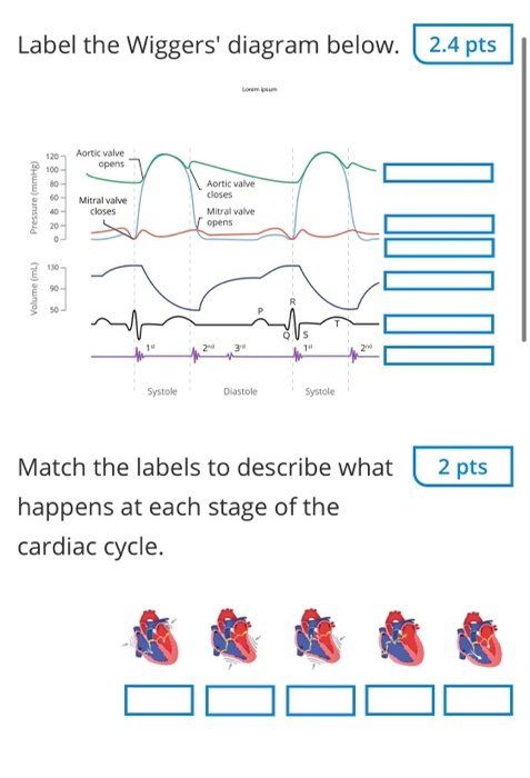
Solved Label the Wiggers' diagram below. 2.4 pts Aortic ...
Cardiac cycle with heart diastole and systole process labeled outline diagram. Illustration about circulation, medicine, artery, care, cardiac, pumping, medical, system - 221540249

Cardiac Cycle | Anatomy and Physiology II
1 Cardiovascular System Components of the Cardiovascular System • consists of the heart plus all the blood vessels • transports blood to all parts of the body in two 'circulations': pulmonary (lungs) & systemic (the rest of the body) • responsible for the flow of blood, nutrients, oxygen and other gases, and hormones to and from cells • about 2,000 gallons (7,572 liters) of blood ...
Heart Anatomy: Labeled Diagram, Structures, Blood Flow ...
Cardiac Cycle Diagram The diagram below represents the different phases of the cardiac cycle. The atrial systole, ventricular diastole, ventricular systole and ventricular diastole are clearly mentioned in the cardiac cycle diagram given below. Cardiac Cycle Physiology The human heart consists of four chambers, comprising left and right halves.
The Cardiac Cycle stock vector. Illustration of muscle ...
The Cardiac Cycle · Filling Phase. The ventricles are filled with blood in two stages – diastole (heart relaxation) and atrial systole (contraction of the atria) ...

Cardiac cycle - Wikipedia
The cardiac cycle - The "Wiggers diagram" Prof. J Swanevelder 4 - 5 Differences in timing between left and right sides of the heart Although the sequence of events on each side of the heart is similar, events occur asynchronously. This disparity in timing reflects differences in anatomy and working pressures between left and right
Solved Describe the cardiac cycle by using the diagrams ...
This chapter is basically a set of long, enormously elaborate footnotes to the Wiggers Diagram, which describes the timing of pressure and ...

Cardiac Cycle - an overview | ScienceDirect Topics
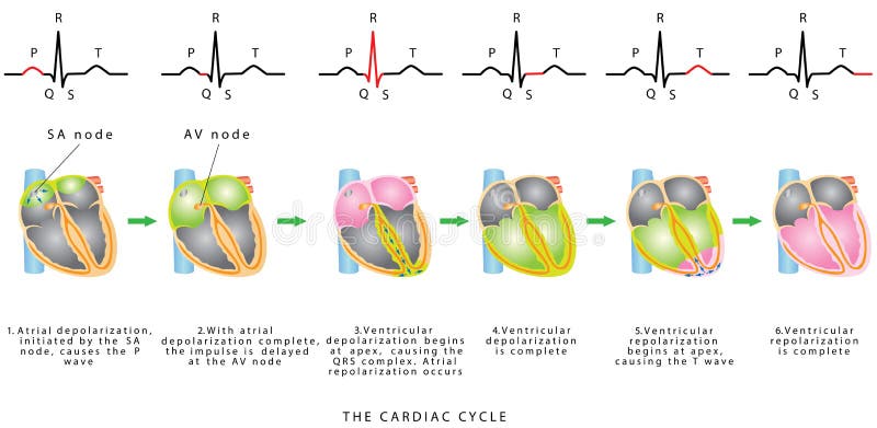
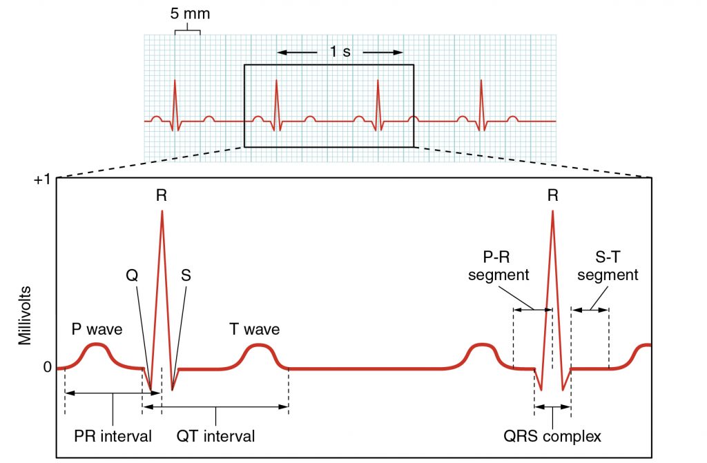
EKG or ECG waveform parts are explained clearly to make EKG interpretation easy. Learn the meaning of each component of an EKG wave with this step-by-step labeled diagram of the conduction system of the heart. Provides information on atrial depolarization and the P wave, ventricular depolarization a
Phases of the Cardiac Cycle
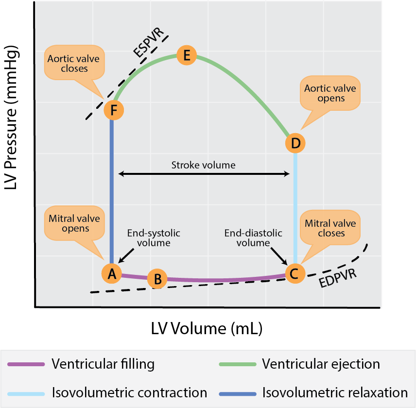
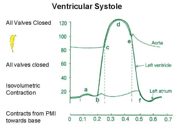
A Wiggers diagram shows the changes in ventricular pressure and volume during the cardiac cycle. Often these diagrams also include changes in aortic and atrial pressures, the EKG, and heart sounds. Diastole starts with the closing of the aortic valve (the second heart sound).

The Cardiac Cycle | Deranged Physiology
Diagram: Blood flow through the heart, cardiac circulation pathway steps, and cardiac anatomy and structures. Blue arrows (deoxygenated blood); Red arrows (oxygenated blood). Now that we have a good understanding of the blood flow through the heart using the cartoon diagrams, we can apply it to a more realistic image of the heart.
Flat Infographics Heart Anatomy Description Cardiac Stock ...
The cardiac cycle has 2 main phases, systole and diastole, defined by whether the heart is depolarized and contracting vs repolarized and relaxed. This post will walk you through one cycle of the heart using a step-by-step diagram that will provide you with easy notes and a simple explanation of cardiac physiology.
Assessment of Cardiovascular Function – A Mixed Course-Based ...
The cardiac cycle is defined as a sequence of alternating contraction and relaxation of the atria and ventricles in order to pump blood throughout the body. It starts at the beginning of one heartbeat and ends at the beginning of another. The process begins as early as the 4th gestational week when the heart first begins contracting.. Each cardiac cycle has a diastolic phase (also called ...
:max_bytes(150000):strip_icc()/heart_electrical_system-597907ca03f4020010e78125.jpg)
Phases of the Cardiac Cycle When the Heart Beats

Left Ventricular Pressure & Volume Changes (Wigger's Diagram ...

Exam 3-Cardiac cycle Diagram | Quizlet

CV Physiology | Cardiac Cycle
LC: Label the heart and describe the cardiac cycle
The Cardiac Cycle The Heart Wiggers Diagram Walk

Cardiac Cycle Phases & Diagram | What is the Cardiac Cycle? Video
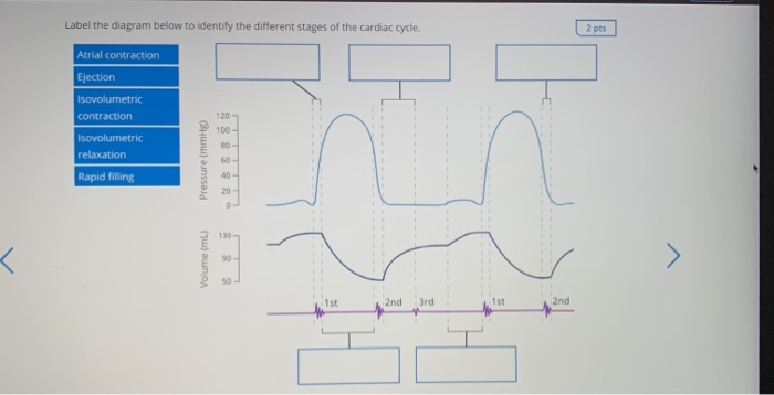
Solved Label the diagram below to identify the different ...

The Cardiac Cycle | Deranged Physiology
Cardiac cycle phases: Definition, systole and diastole | Kenhub

Cardiac Cycle Phases and Blood Flow: Step-By-Step Heart ...

Label this: stages of the cardiac cycle Diagram | Quizlet

The Wright table of the cardiac cycle: a stand-alone ...

Cardiac Cycle | BioNinja
The heart cycle: The mammalian heart & cardiac cycle ...

0714 Cardiac Cycle Medical Images For PowerPoint | PowerPoint ...

Left ventricular diastolic function – ECG & ECHO
.png)
Cardiac Cycle Quiz: How Well You Know? - ProProfs Quiz
Phases of the Cardiac Cycle - Diastole, Atrial Systole and ...
Cardiac system 1: anatomy and physiology | Nursing Times
0 Response to "0 cardiac cycle diagram labeled"
Post a Comment