42 Drag The Labels Onto The Diagram Of Muscle Spindle Function.
Chapter 8 Homework - Amazonassignments.com Chromosomes condense and are attached to spindle fibers. Cytokinesis. ... During _____, the cell carries out its normal functions and the chromosomes are thinly spread out throughout the nucleus. ... Drag the labels onto the diagram to identify the stages of the cell cycle. A&P 1 (Histology) Flashcards - Quizlet Drag the labels to the appropriate location in the figure. Identify the type and tissue shown Cardiac Muscle Tissue Identify the type and tissue shown Loose Connective Tissue Drag the labels onto the diagram to identify the tissues and structures Identify the structure at the tip of the pointer. Nucleus Identify the ground substance for this tissue
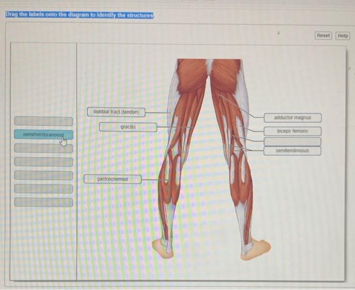
Bio 2331 Prelab 6 Muscles Part 1.pdf - 2/10/22, 10:55 PM ... 2/10/22, 10:55 PM Bio 2331 Prelab 6 Muscles Part 1 Art-labeling Activity: Classification of Muscles by Function Part A Drag the labels to the appropriate location in the figure. ANSWER: Lab Manual Exercise 11 From the Book Pre-lab Quiz Question 3 Part A When a muscle contracts to cause an action at a joint, the origin is the moving attachment ...
Drag the labels onto the diagram of muscle spindle function.
PDF CHAPTER 4: TISSUES - Warner Pacific University Function: Transmit electrical signals from sensory receptors and to effectors (muscles and glands) which control their activity. Location: Brain, spinal cord, and nerves. Description: Neurons are branching cells; cell processes that may be quite long extend from the nucleus-containing cell body; also contributing to nervous tissue Chapter 13 Physiology Mastering A&P Flashcards | Quizlet In the last part of this coaching activity you will turn your attention to the crossed extensor reflex. You will first order the events that take place during this reflex. Then you will identify the type (s) of synapses found in this reflex arc. Finally, you will test your knowledge by applying what you have learned to a different/new reflex. Drag The Labels Onto The Diagram To Identify The Parts Of ... Drag the correct description under each cell structure to identify the role it plays in the cell. This problem has been solved. Drag the labels onto the diagram to identify the parts of the cell. Drag the labels onto the diagram to identify the mechanisms involved in the transport of carbon d. Part a animal cell structure drag the labels onto ...
Drag the labels onto the diagram of muscle spindle function.. 40 label the structures involved in muscle spindle function. Transcribed image text: Drag the labels onto the diagram of muscle spindle function. Reset Muscle shortens Strelch receptors in the spinde are activated throughout the contraction Muscle length Gamma mator neurons activate intrafusal fibers Intrafusal fibers do not slacken so firing rate remains constant. bio 141 mastering ch 7 12.1-12.2 Flashcards | Quizlet Part A - Animal cell structures and functionsTo understand how cells function as the fundamental unit of life, you must first become familiar with the individual roles of the cellular structures and organelles.Drag the labels on the left onto the diagram of the animal cell to correctly identify the function performed by each cellular structure. Solved Part A Drag the labels onto the diagram to identify ... Transcribed image text: Part A Drag the labels onto the diagram to identify the parts of a knee-jerk reflex. THE PATELLAR TENDON (K Monosynaptic activation of somatic motor neuron Sensory neuron an action potential Muscle spindle stretched and Effector contracts stretch-sensitive neurons are activated Inhibited motor neuron does not activate antagonistic muscle Response extension of the leg ... 10.2 Skeletal Muscle - Anatomy & Physiology Figure 10.2.2 - Muscle Fiber: A skeletal muscle fiber is surrounded by a plasma membrane called the sarcolemma, which contains sarcoplasm, the cytoplasm of muscle cells. A muscle fiber is composed of many myofibrils, which contain sarcomeres with light and dark regions that give the cell its striated appearance.
Solved Drag the labels onto the diagram of muscle spindle ... Transcribed image text: Drag the labels onto the diagram of muscle spindle function. Reset Muscle shortens Strelch receptors in the spinde are activated throughout the contraction Muscle length Gamma mator neurons activate intrafusal fibers Intrafusal fibers do not slacken so firing rate remains constant. what is the function of a muscle spindle? Seleccionar página. what is the function of a muscle spindle? por | kiasuparents o-level 2021 | kiasuparents o-level 2021 what is the function of a muscle spindle? is south carolina under a state of emergency. what is the function of a muscle spindle? what is the function of a muscle spindle? Drag the labels on the left onto the diagram of the animal ... Drag the labels on the left onto the diagram of the animal cell to correctly identify the function performed by each cellular structure. Hint 1. Structure and function of cell organelles The structure of each organelle in a eukaryotic cell makes it very well-suited for the task it performs. Some examples are described here.
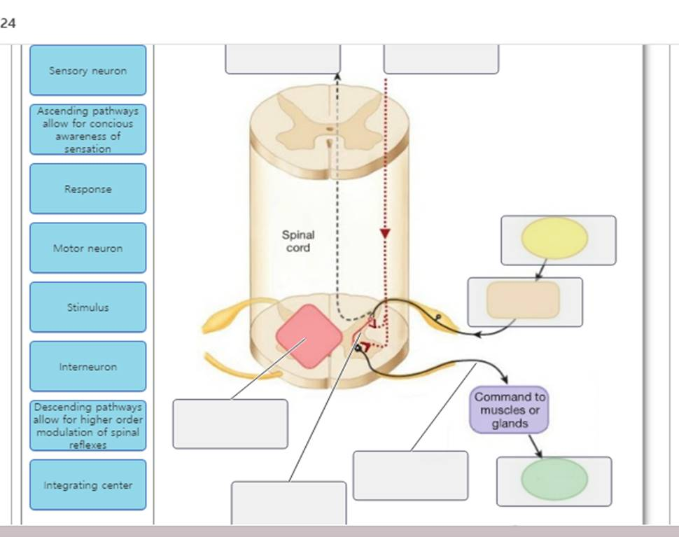
Chapter 13.pdf - Chapter 13 Chapter 13 Due 12:00am on ... Part A Drag the labels onto the diagram of muscle spindle function. ANSWER: Correct Essentials Figure: The Crossed Extensor Reflex View figure 13.1 in greater detail Help Reset Gamma motor neurons activate intrafusal fibers Stretch receptors in the spindle are activated throughout the contraction Alpha motor neurons activate extrafusal muscle ... Chapter 8 Homework - Free Essay Examples Database Chromosomes condense and are attached to spindle fibers. Cytokinesis. ... During _____, the cell carries out its normal functions and the chromosomes are thinly spread out throughout the nucleus. ... Drag the labels onto the diagram to identify the stages of the cell cycle. Muscles Of The Eye Superior View Diagram - Studying Diagrams They are the Superior and Inferior Rectus Muscles. Drag the labels onto the diagram to identify the muscles of facial expression lateral view. Medial View Right Eye. The position of the eye at the time of muscle contraction is what determines. The superior oblique also moves the eye downward. Drag the labels onto the diagram to identify the parts of a ... Jan 02, 2022 · Label the components of a knee-jerk reflex Part A Drag the labels onto the diagram to identify the parts of a knee-jerk reflex. THE PATELLAR TENDON Inhibited motor neuron does not activate antagonistic muscle Response extension of the leg Muscle spindle stretched and nosyn tic activation of somatic motor stretch-sensitive neurons are neuron Activated conducts an actioninhibitory Sensory neuron ...
Reflex Physiology. automatic, unconscious to changes, either ...
what is the function of a muscle spindle? Feb 17, 2022 · Uma senha será enviada por e-mail para você. study on generation z travellers european travel commission Minilua. regency healthcare careers; triathlon taren heart rate spreadsheet
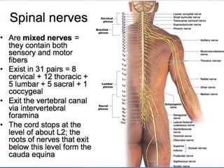
The Nervous System: The Spinal Cord and Spinal Nerves
Physiology Chapter 12 Assignment ML Flashcards | Quizlet Gravity Created by mirandaluke Terms in this set (70) Drag the labels onto the diagram to label the steps of smooth muscle activation and deactivation. Action potential propagation in a skeletal muscle fiber ceases when acetylcholine is removed from the synaptic cleft.
SENSE ORGANS HANDOUT
NDSU Human Anat I- Exam 2 Flashcards - Quizlet Drag the resting membrane determinants to their appropriate locations in the figure. Answers may be used once, or not at all. Drag the appropriate labels to their targets. Note that pink labels should go in pink targets, blue labels should go in blue targets, and green labels should go in green targets.

Lab Activity chapter 21.pdf - 4/17/2020 Lab Activity chapter ...
Drag The Labels Onto The Diagram Of Muscle Spindle ... If you searching to evaluate drag the labels onto the diagram of muscle spindle function drag the labels onto the diagram of muscle spindle function price. Buy online keeping the car safe transaction. This item is incredibly nice product. When an antigen is bound to a class ii mhc protein it can activate a cell.

Interactions of Skeletal Muscles | Lifetime Fitness and Wellness
Drag The Labels Onto The Diagram To Identify The Parts Of ... Drag the correct description under each cell structure to identify the role it plays in the cell. This problem has been solved. Drag the labels onto the diagram to identify the parts of the cell. Drag the labels onto the diagram to identify the mechanisms involved in the transport of carbon d. Part a animal cell structure drag the labels onto ...
Anatomy and Physiology Lab I” on OpenALG
Chapter 13 Physiology Mastering A&P Flashcards | Quizlet In the last part of this coaching activity you will turn your attention to the crossed extensor reflex. You will first order the events that take place during this reflex. Then you will identify the type (s) of synapses found in this reflex arc. Finally, you will test your knowledge by applying what you have learned to a different/new reflex.

Label the components of a knee-jerk reflex Part A Drag the labels
PDF CHAPTER 4: TISSUES - Warner Pacific University Function: Transmit electrical signals from sensory receptors and to effectors (muscles and glands) which control their activity. Location: Brain, spinal cord, and nerves. Description: Neurons are branching cells; cell processes that may be quite long extend from the nucleus-containing cell body; also contributing to nervous tissue
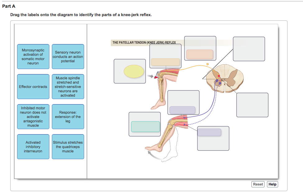
Solved Part A Drag the labels onto the diagram to identify ...
Kinesiology

A&P2 Lab 7 HW, A&P2 Lab 6 HW, A&P 2 Lab 5 HW, A&P2 Lab 7 PP ...

Bones of the Upper Limb | Anatomy and Physiology I

Overview of the Muscular System | Boundless Anatomy and ...
Muscles and Muscle Tissue

A&P2 Lab - Lesson 1 Flashcards | Quizlet

Lab Activity chapter 21.pdf - 4/17/2020 Lab Activity chapter ...
Chapter13 15(one section)

Bones of the Upper Limb | Anatomy and Physiology I

Taste bud - Wikipedia

JaypeeDigital | eBook Reader

JaypeeDigital | eBook Reader
.png)
The Cell Cycle, Mitosis And Meiosis - ProProfs Quiz
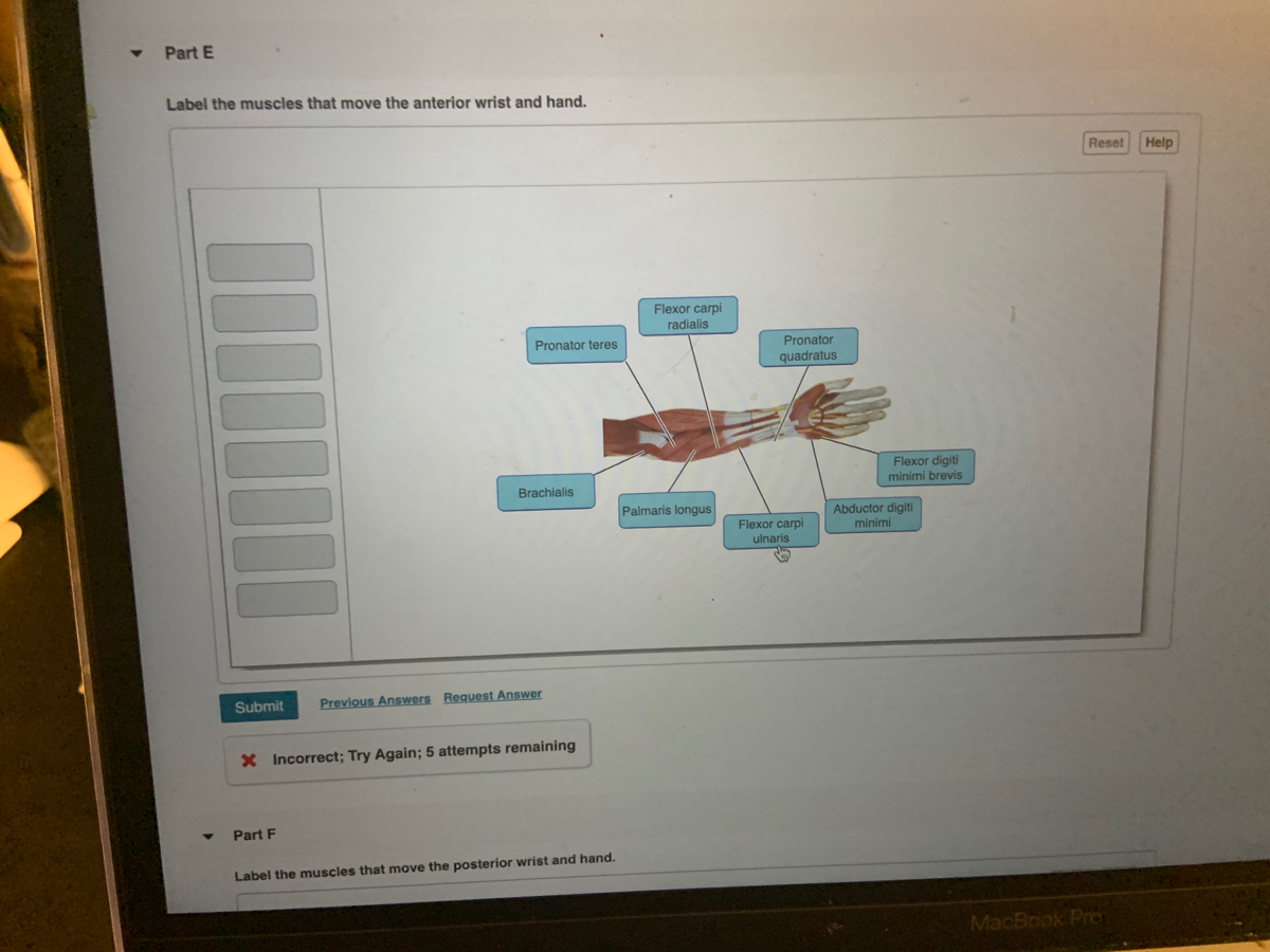
Answered: Label the muscles that move the… | bartleby
Solved Drag the labels onto the figure to identify the parts ...
Anatomy and Physiology Lab I” on OpenALG

The Glossary of Prosthodontic Terms - Journal of Prosthetic ...
SENSE ORGANS HANDOUT
Solved Drag the labels onto the diagram to identify the ...
Ascending tracts of the spinal cord: Anatomy | Kenhub

Lab Activity chapter 21.pdf - 4/17/2020 Lab Activity chapter ...

Chapter 47 - Sensory Perception - BIO 140 - Human Biology I ...

Reflexes | Anatomy and Physiology I

Drag the labels to identify the classes of lymphocytes. Hasat ...
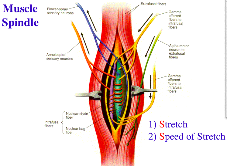
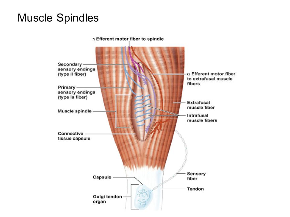
Muscle Spindle and Golgi Tendon Organ

Diagrammatic representation of muscle spindle. | Download ...

BIO23 F19-S20 Complete Course Guide by Human Anatomy - Issuu

Week 6: Muscle Physiology Flashcards & Practice Test | Quizlet

BIO 360: Animal Physiology - MasteringPhysiology Pre-class ...

Lab Activity chapter 21.pdf - 4/17/2020 Lab Activity chapter ...
Neural Control of Human Movement

1.4 The Somatic Nervous System – Neuroscience: Canadian 1st ...
Treatment Approaches and Options Michael G. Kaiser Regis W ...

Lab Activity chapter 21.pdf - 4/17/2020 Lab Activity chapter ...
0 Response to "42 Drag The Labels Onto The Diagram Of Muscle Spindle Function."
Post a Comment