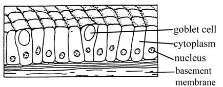
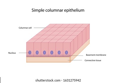
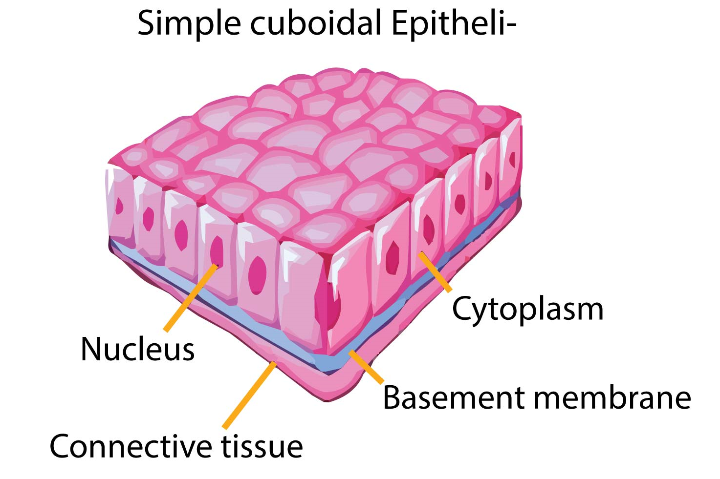
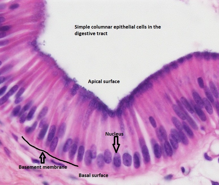
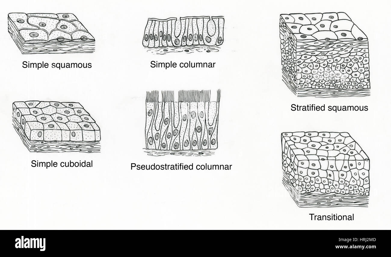
44 simple columnar epithelium labeled diagram
courses.lumenlearning.com › boundless-ap › chapterLayers of the Alimentary Canal | Boundless Anatomy and Physiology The most variation is seen in the epithelium tissue layer of the mucosa. In the esophagus, the epithelium is stratified, squamous, and non-keratinizing, for protective purposes. In the stomach. the epithelium is simple columnar, and is organized into gastric pits and glands to deal with secretion. Simple Columnar Epithelium - Definition & Function ... Simple columnar epithelia are tissues made of a single layer of long epithelial cells that are often seen in regions where absorption and secretion are important features. The cells of this epithelium are arranged in a neat row with the nuclei at the same level, near the basal end. In a cross-section of the organ, these cells appear like thin ...
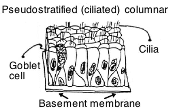
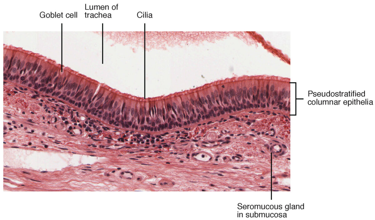
Epithelia: The Histology Guide Simple secretory columnar epithelium lines the stomach and uterine cervix.The simple columnar epithelium that lines the intestine also contains a few goblet cells. In histological slides of pseudostratified epithelium, it looks as though some of the cells are not in contact with the basal lamina, and the nuclei are at different levels.

Simple columnar epithelium labeled diagram
histology.medicine.umich.edu › resourcesConnective Tissue and Quiz 1 | histology Look at the areas outlined in the orientation diagram of the trachea and locate the loose, cellular connective tissue within the glands (the "glands" are coiled tubes of columnar epithelial cells; some the epithelial cells are tall and eosinophilic, whereas others are shorter and more basophilic). Simple Columnar Epithelium Labeled Diagram Simple Columnar Epithelium Labeled Diagram. Ciliated columnar epithelium is composed of simple columnar epithelial cells with cilia on their apical This illustration shows a diagram of a goblet cell. These labelled diagrams should closely follow the current Science (simple squamous epithelium). Simple Columnar Epithelium Labeled Diagram and Function ... Simple Columnar Epithelium Labeled Diagram and Function - YouTube. Simple Columnar Epithelium Labeled Diagram and Function. Watch later. Share. Copy link. Info. Shopping. Tap to unmute. If ...
Simple columnar epithelium labeled diagram. Label the diagram of simple squamous epithelium Diagram ... Start studying Label the diagram of simple squamous epithelium. Learn vocabulary, terms, and more with flashcards, games, and other study tools. courses.lumenlearning.com › boundless-ap › chapterThe Male Reproductive System | Boundless Anatomy and Physiology The epithelium of the tubule consists of tall, columnar cells called Sertoli cells. Between the Sertoli cells are spermatogenic cells, which differentiate through meiosis to become sperm cells. There are two types of seminiferous tubules: convoluted, located toward the lateral side, and straight, as the tubule comes medially to form ducts that ... Simple Columnar Epithelium Labeled Diagram Simple Columnar Epithelium: A Labeled Diagram and Functions Epithelium is a tissue that lines the internal surface of the body, as well as the internal organs. Simple epithelium is one of the types of epithelium that is divided into simple columnar epithelium, simple squamous epithelium, and simple cuboidal epithelium. Ciliated epithelium is a thin tissue that has hair-like structures on it. These hairs, called cilia, move back and forth to help move particles out of our body. › 44118315 › The_Biology_of_Cancer(PDF) The Biology of Cancer- R.Weinberg - Academia.edu Academia.edu is a platform for academics to share research papers.
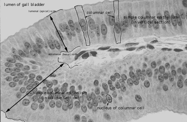
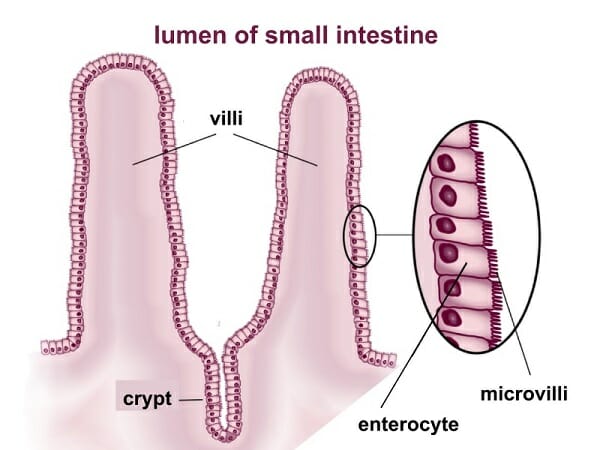
Simple Squamous Epithelium Function: Passage of materials ... Simple Squamous Epithelium Function: Passage of materials by Location: Air sacs of lungs Simple Cuboidal Epithelium Function: Secretion and Absorption Location: Glandular tissue, kidney tubules Simple Columnar Epithelium Function: Absorption Location: Lining of the digestive tract. Simple Squamous Epithelium. Ileum Histology Slide with Labeled Diagram and ... You will find different types of cells that make up the ileum. First, you will find the absorptive columnar epithelium in the mucosal surface of the ileum structure. Again, you will find the glandular epithelium in the intestinal glands of the ileum slide. The surface epithelium (simple columnar epithelium) contains microvilli on their surfaces. study.com › learn › lessonSimple Cuboidal Epithelium Function & Location | What Is ... Simple Cuboidal Epithelium: Labeled Diagram. Simple cuboidal epithelial cells are shaped like cubes, and the nucleus of each cell is large and located close to the center of the cell. This is ... Simple columnar epithelium - Eugraph These absorptive cells are a single layer of columnar cells. (a simple columnar epithelium). Note an oval nucleus in the lower part of each columnar cell. Arrows indicate the base of this simple columnar epithelium sce. The lumen is indicated by lu. The surface area for absorption is increased by projections of the intestinal wall called villi.
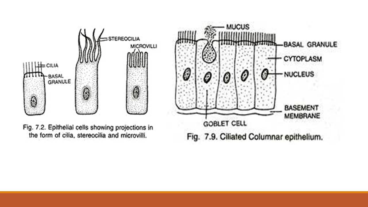
Jejunum Histology Slide with Labeled Diagram and ... The lining epithelium of the tunica mucosa is a simple columnar epithelium with goblet cells. You may also find other different cells in the mucosa of the small intestine like penath cells, enteroendocrine cells, and others. The number of the plica circularis and villi mary varies in the different parts of an animal's small intestine. Simple epithelium: Location, function, structure | Kenhub Ciliated columnar epithelium is therefore found in the respiratory tract where mucous and air are pushed away to clear the respiratory tract. Other areas where ciliated columnar epithelium is found are the fallopian tubes, the uterus, and the central canal of the spinal cord. In the female reproductive system, the ciliated columnar epithelium lines the lumen of the uterine tube and the movements generated by the cilia propel the egg towards the uterus. PDF Intestine Cell Diagram - yearbook2017.psg.fr epithelium is a tissue that lines the internal surface of the body as well as the internal organs simple epithelium is one of the types of epithelium that is divided into simple columnar epithelium simple squamous epithelium and simple cuboidal epithelium bodytomy provides a labeled diagram to help you understand the structure and function of ... Simple squamous epithelium - Eugraph Epithelial tissue: Simple squamous epithelium Example: A simple squamous epithelium forms the outer wall of the glomerular capsule. The structure highlighted with normal color is, in three-dimensions, a sphere composed of a thin outer wall of cells, a space that contains fluid, and an inner region of cells.
Simple cuboidal epithelium Simple cuboidal epithelium (400X) Kidney cortex The arrow is pointing to a cross section of a tube made of simple cuboidal epithelium. Start at the point of the arrow and follow the nuclei in a circle until you get back to the arrow. There are several other tubes shown in cross section on this image.
anatomylearner.com › epithelial-tissueEpithelial Tissue - Types, Location, Examples and Histology ... Apr 04, 2021 · Functions of simple columnar epithelium and their location. Absorption and secretion are the primary functions of simple columnar epithelium. You will find simple columnar epithelium in the following organs or structures of an animal’s body. #1. Simple ciliated columnar epithelium in the respiratory tract, uterine tube #2.
PDF Basic Histo diagrams labelled in colour - 2005 SIMPLE EPITHELIA EPITHELIUM: simple cuboidal TISSUE / ORGAN: thyroid EPITHELIUM: simple squamous TISSUES / ORGANS: 1 endothelium lining blood vessel 2 mesothelium of serosa covering lung EPITHELIUM: simple columnar TISSUE / ORGAN: gall bladder EPITHELIUM: pseudostratified columnar (respiratory epithelium) MORE FULLY: pseudostratified ciliated
Pseudostratified Columnar Epithelium | Histology, Anatomy ... The epithelium, the outermost layer of the body surface, is consist of a layer (or layers) of similar cells that are bound closely together. Learn about the pseudostratified columnar epithelium (anatomy, types & functions) and find out what makes it unique from other epithelia found in the body.
Stratified Cuboidal Epithelium - Definition and Function ... Stratified Cuboidal Epithelium Definition. Stratified cuboidal epithelium is a type of epithelial tissue found mainly in glands, which specialize in selective absorption and secretion by the gland into blood or lymph vessels. In general, epithelial tissue is any group of cells lining a body cavity or body surface. Stratified cuboidal epithelium describes an epithelial tissue with two aspects.
Simple Squamous Epithelium: Location and Diagram - Video ... The simple squamous epithelium is different from other types of epithelial tissue such as simple cuboidal, simple columnar, and stratified squamous epithelium in that it is only made of one layer ...
Epithelial Tissue: Structure with Diagram, Function, Types ... Simple Epithelium- it is composed of one layer of a cell and mostly has a secretory or an absorptive function. Compound (Stratified) Epithelium- it is made up of two or more than two layers of cells and mostly has a protective function. The glandular epithelium is made up of cuboidal or columnar cells. They are specialised for secretion.
Simple Columnar Epithelium Diagram - Quizlet The surface area is increased with microvilli and its function is to move materials, secrete fluids, and absorb nutrients. What are goblet cells and their functions? Goblet cells are glandular simple columnar epithelial cells whose function is to produce and secrete mucus in order to protect the mucous membranes where they are found.
› 37006818 › Junqueiras_Basic(PDF) Junqueira's Basic Histology Text and Atlas, 14th ... Junqueira's Basic Histology Text and Atlas, 14th Edition
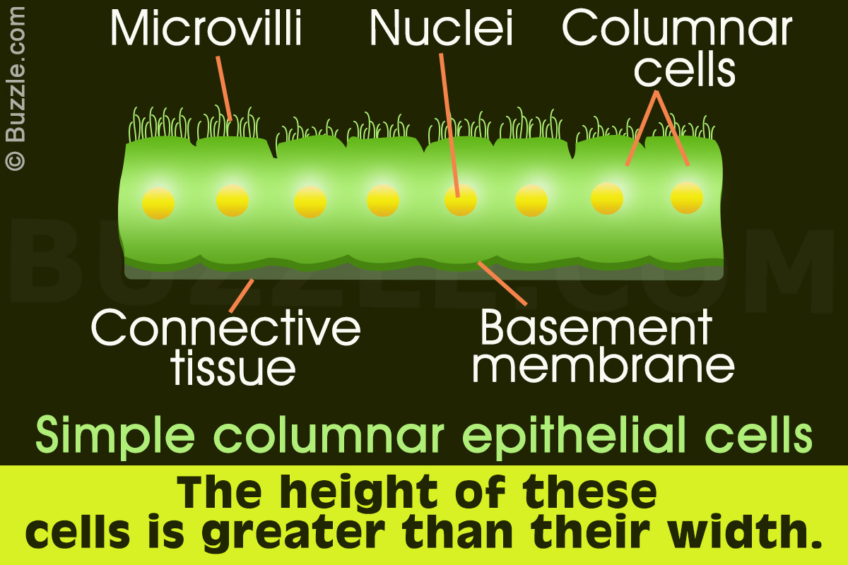
Simple Columnar Epithelium: A Labeled Diagram and ... Simple Columnar Epithelium: A Labeled Diagram and Functions Epithelium is a tissue that lines the internal surface of the body, as well as the internal organs. Simple epithelium is one of the types of epithelium that is divided into simple columnar epithelium, simple squamous epithelium, and simple cuboidal epithelium.
Articles - Page 4 of 20 - Bodytomy Simple Columnar Epithelium: A Labeled Diagram and Functions Epithelium is a tissue that lines the internal surface of the body, as well as the internal organs. Simple epithelium is one of the types of Understanding Negative and Positive Feedback in Homeostasis Made Easy
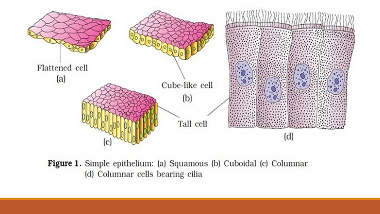
Describe various types of epithelial tissues with the help ... (iii) Simple columnar epithelium: It consists of a single layer of tall, slender cells with their nuclei present at the base of the cells. They may bear microvilli on the free surfaces. Columnar epithelium forms the lining of the stomach and intestines and is involved in the function of secretion and absorption.
Ileum: Anatomy, histology, composition, functions - Kenhub It is made up of simple squamous epithelium and a connective tissue layer underneath (lamina propria serosae). A characteristic feature of the ileum is the Peyer's patches lying in the lamina propria of mucosa and in the submucosa. It is an important part of the GALT (gut-associated lymphoid tissue). One patch is around 2 to 5 centimeters long and consists of about 300 aggregated lymphoid follicles and the parafollicular lymphoid tissue.
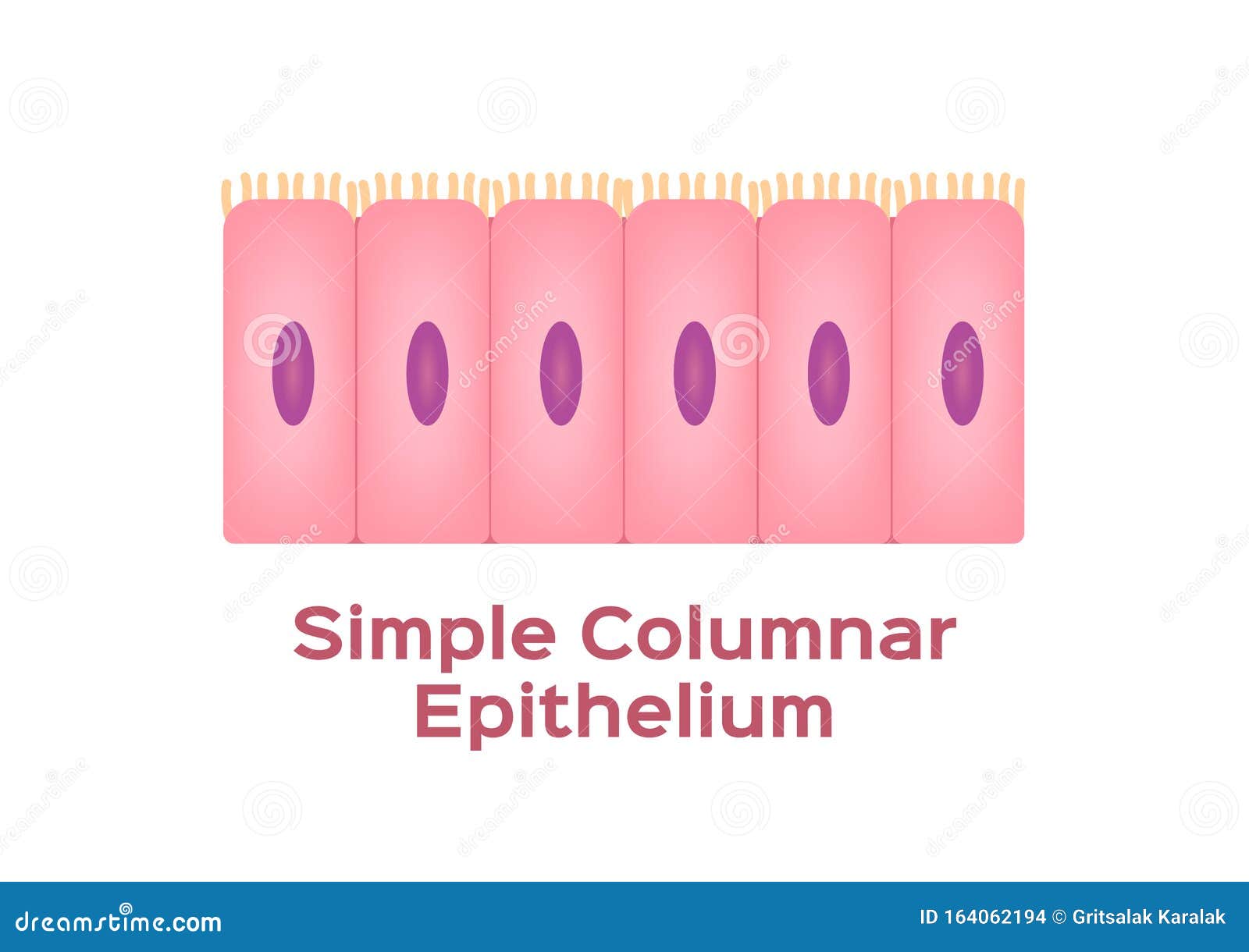
Simple columnar epithelium- structure, functions, examples The simple columnar epithelium is a type of epithelium that is formed of a single layer of long, elongated cells mostly in areas where absorption and secretion are the main functions. Like cuboidal epithelium, the cells in the columnar epithelium are also modified to suit the function and structure of the organ better.
Simple Columnar Epithelium Labeled Diagram and Function ... Simple Columnar Epithelium Labeled Diagram and Function - YouTube. Simple Columnar Epithelium Labeled Diagram and Function. Watch later. Share. Copy link. Info. Shopping. Tap to unmute. If ...
Simple Columnar Epithelium Labeled Diagram Simple Columnar Epithelium Labeled Diagram. Ciliated columnar epithelium is composed of simple columnar epithelial cells with cilia on their apical This illustration shows a diagram of a goblet cell. These labelled diagrams should closely follow the current Science (simple squamous epithelium).
histology.medicine.umich.edu › resourcesConnective Tissue and Quiz 1 | histology Look at the areas outlined in the orientation diagram of the trachea and locate the loose, cellular connective tissue within the glands (the "glands" are coiled tubes of columnar epithelial cells; some the epithelial cells are tall and eosinophilic, whereas others are shorter and more basophilic).








(253).jpg)






0 Response to "44 simple columnar epithelium labeled diagram"
Post a Comment