45 human heart blood flow diagram
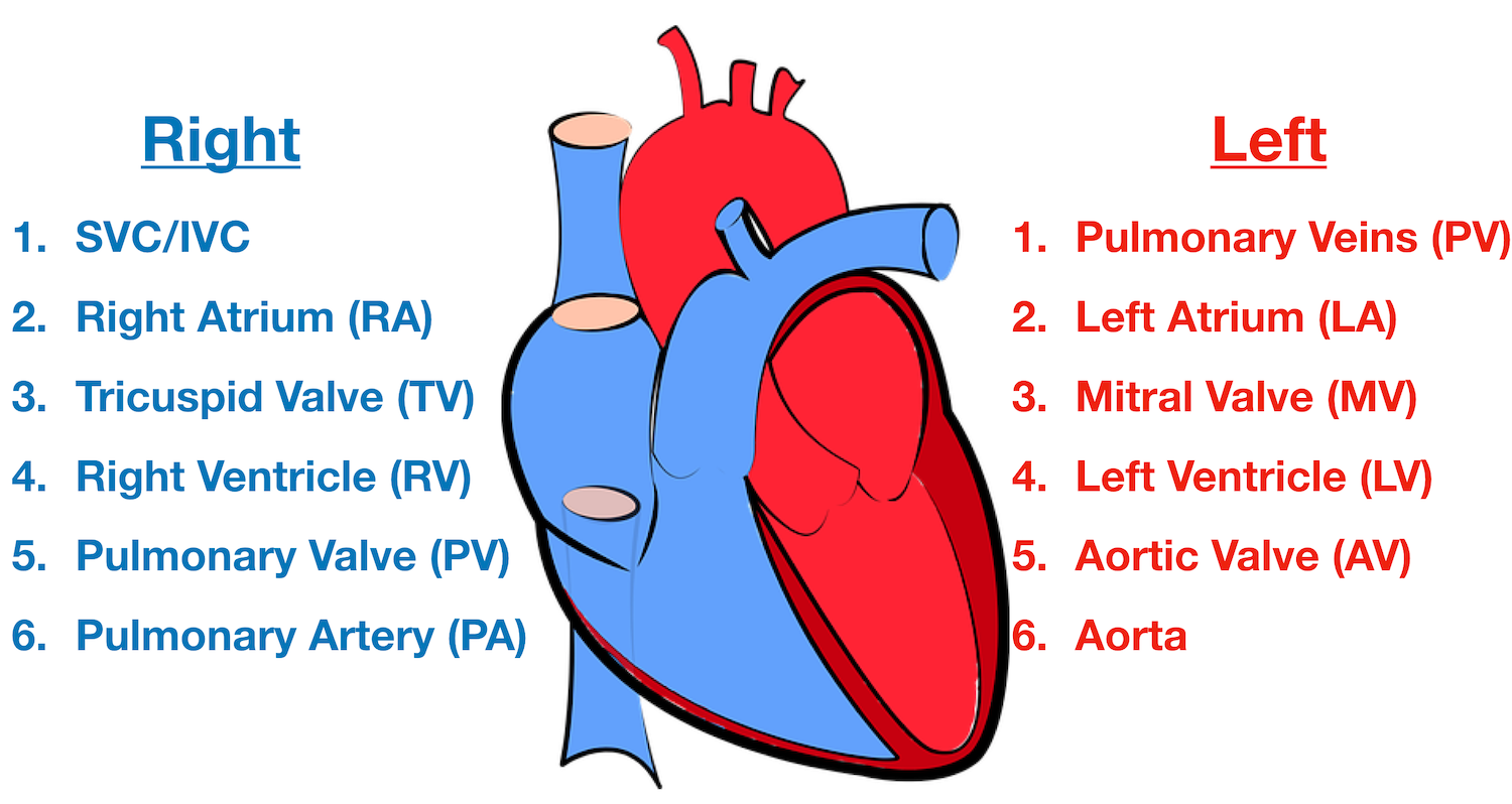
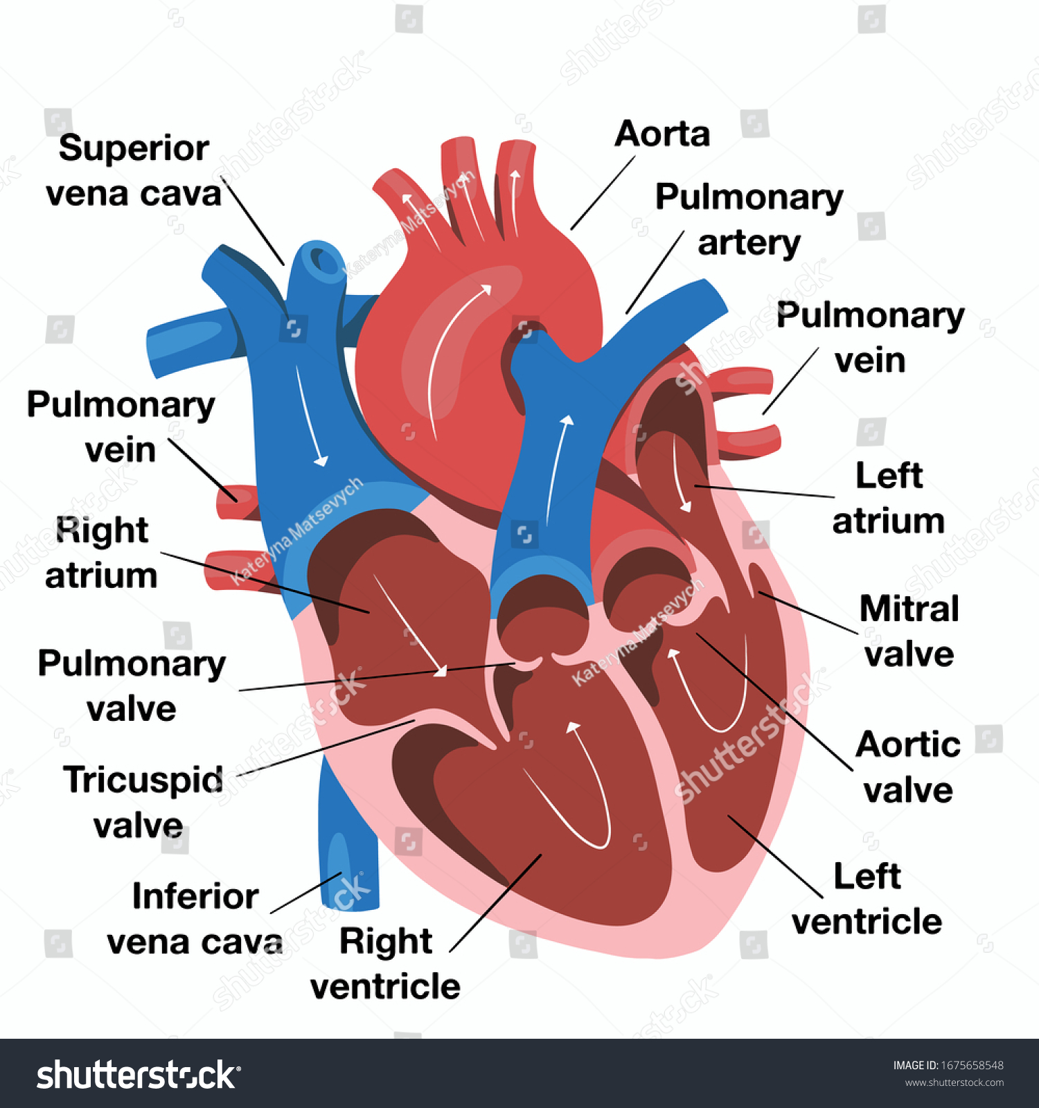
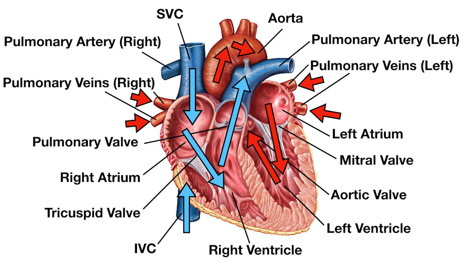
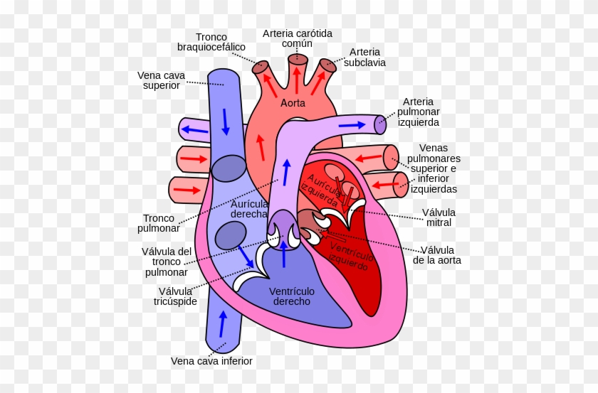
The mitral and tricuspid valves are atrioventricular (AV) valves located between the atria and the ventricles. Two semilunar (crescent-shaped, like a half-moon) valves, the aortic and pulmonary valves, are located in the arteries leaving the heart. The mitral valve is the only bicuspid valve in the human heart. - Human Heart Diagram. Theyre just there to square. Label the right and left atria as RA and LA Label the right and left ventricle as RV and LV Remember that you are looking at an illustration of someone elses heart. ... Blood flow throughout the body is summarized. The first two rounds of this heart are just a simple granny square.
Opening a new path to the effective delivery of biological factors (e.g., stem cells, growth factors, drugs, small interfering RNA (siRNA), and microRNA) to injured tissue sites, engineered biodegradable scaffolds are capable of creating suitable extracellular matrices (ECMs) for implanted cells, thus facilitating and enhancing tissue regeneration.

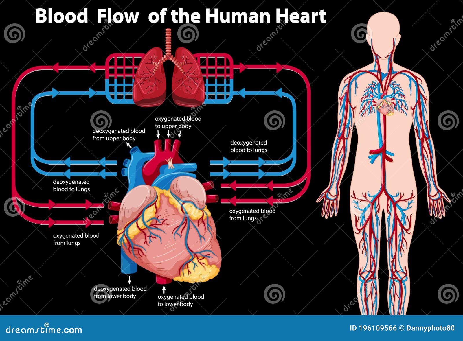
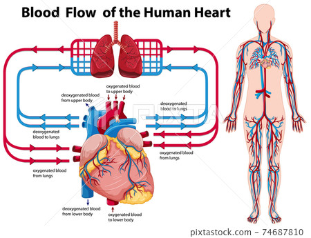
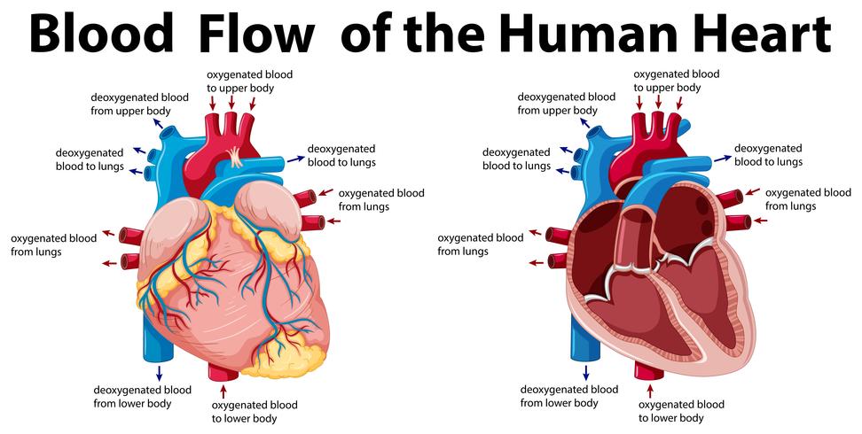
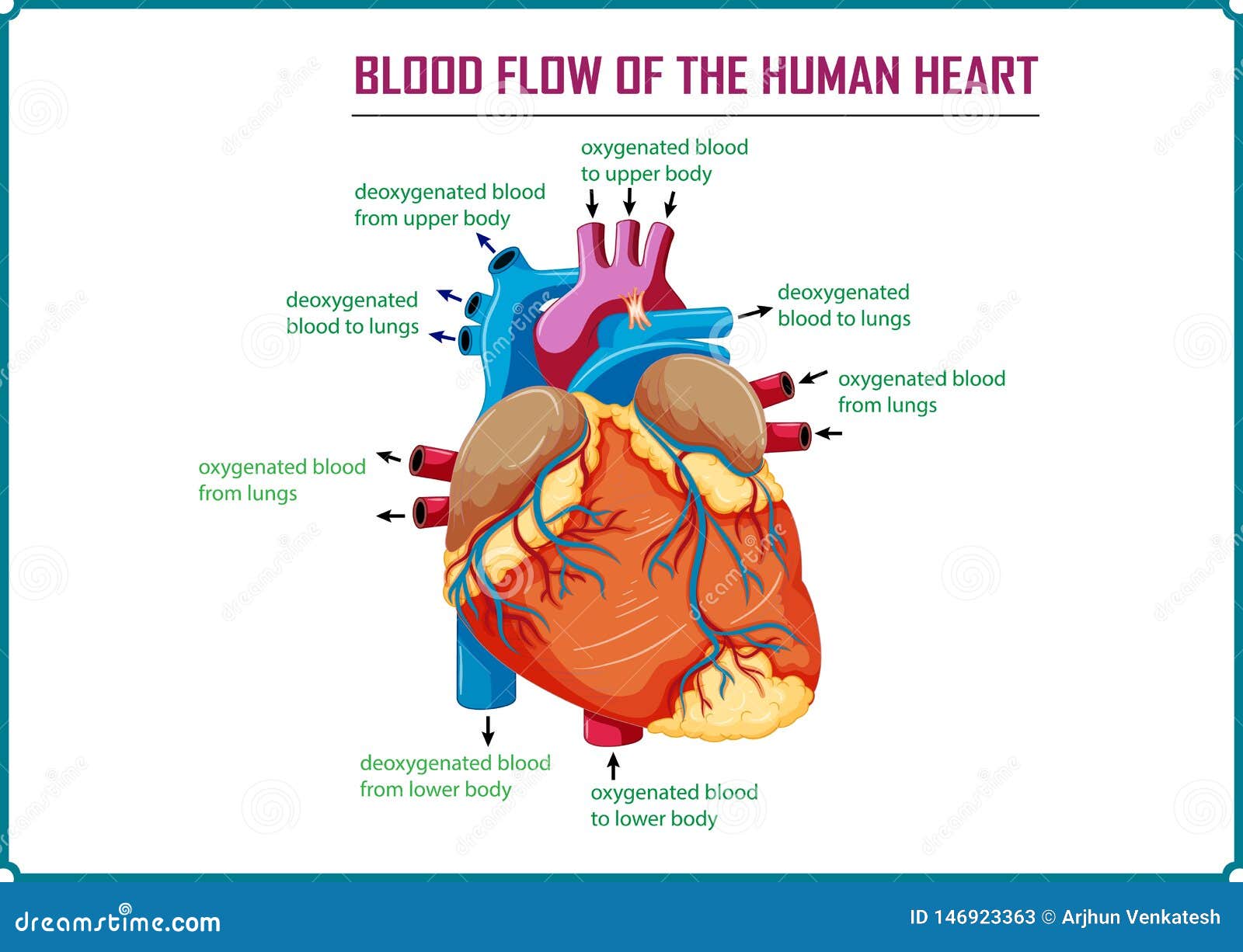
Human heart blood flow diagram
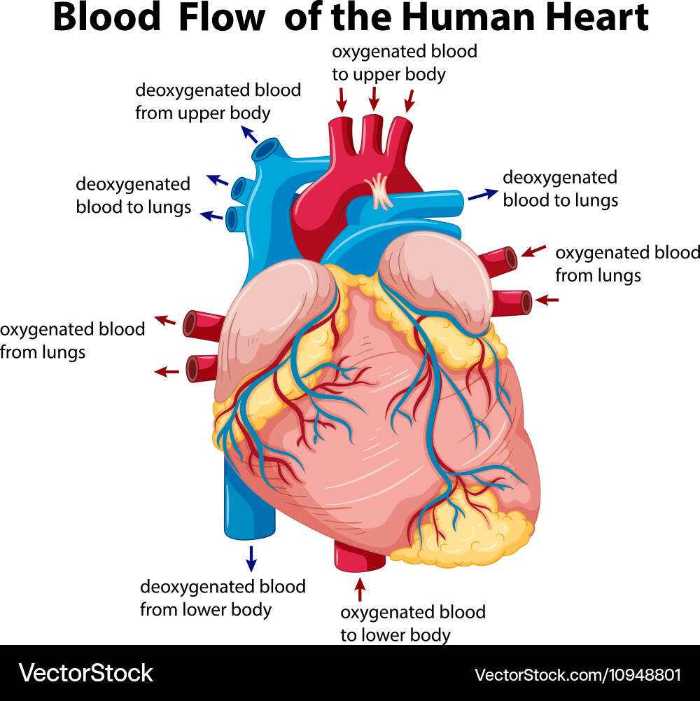
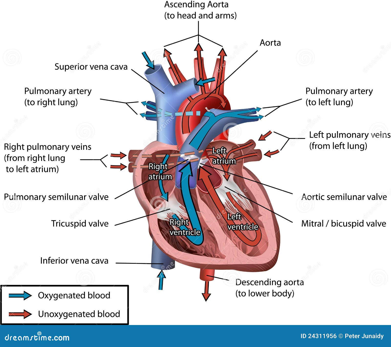
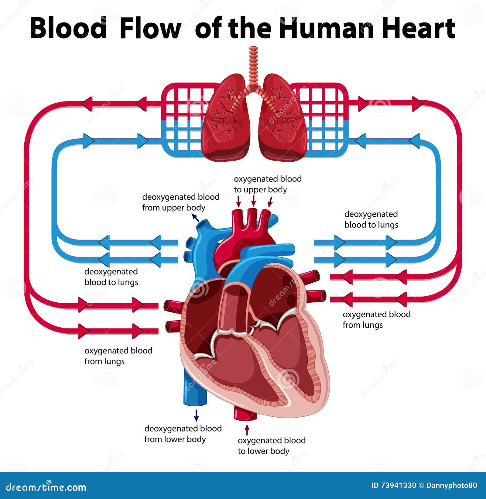
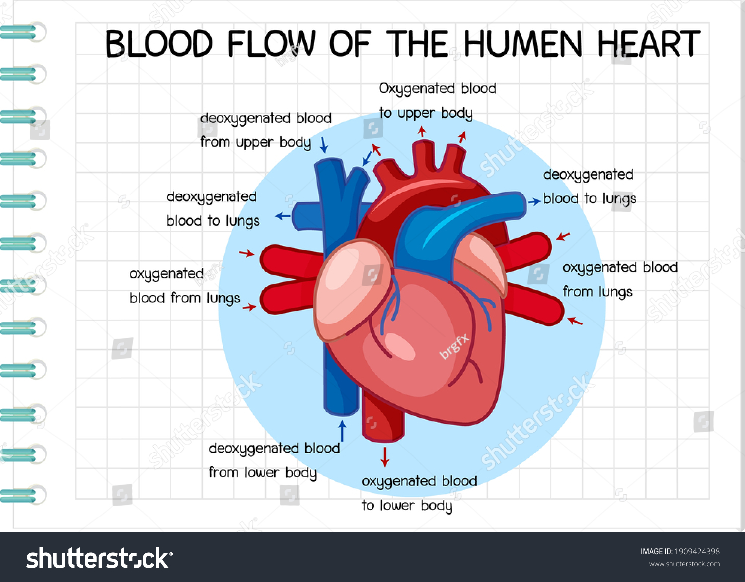
Oxygenated blood flows back to the heart through the pulmonary vein, back into your left atrium. From the atrium, oxygen-rich blood empties into the left ventricle through another valve (mitral valve). The left ventricle is a powerful muscle that contracts to pump blood into the systemic circulation. Blood passes through the aortic valve and ... Arteries Diagram Heart : Human Blood Vessels Anatomy - YouTube - If you experience these or similar symptoms, call yo. Get link; Facebook; Twitter; Pinterest; Email; ... The intercostal artery refers to the set of blood vessels that direct blood flow to an area within the ribs known as the intercostal space. The heart is a mostly hollow ... White blood cells Red blood cells Antibodies Plasma. Question 5 (Multiple Choice Worth 2 points) (06.05 MC)This is a two-part diagram showing maternal circulation on the left and fetal circulation on the right. Letter A indicates a large crescent-shaped organ that contains blood from the maternal circulation and blood from the fetal circulation.
Human heart blood flow diagram. Figure 1 shows the simplest of two component phase diagram s. The components are A and B, and the possible phases are pure crystals of A, pure crystals of B, and liquid with compositions ranging between pure A and pure B. Compositions are plotted across the bottom of the diagram. The normal human body temperature is often stated as 36.5-37.5 °C (97.7-99.5 °F). ... Vasodilation increases blood flow to the skin, resulting in heat being lost to the environment. This produces the effect of feeling warm, when ... which allows the heart to be stopped and blood pressure to be lowered to zero, for the treatment of ... IBM's DiscoveryLink for the life sciences is one of the best known. 12 The Kleisli system provides an internal nested complex data model and a high-power query and transformation language for data integration. 13 K2 shares many design principles with Kleisli in supporting a complex data model, but adopts more object-oriented features. 14 OPM supports a rich object model and a … The most common side effects of VENCLEXTA when used in combination with obinutuzumab or rituximab or alone in people with CLL or SLL include low white blood cell counts; low platelet counts; low red blood cell counts; diarrhea; nausea; upper respiratory tract infection; cough; muscle and joint pain; tiredness; and swelling of your arms, legs ...
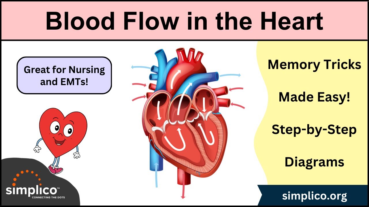
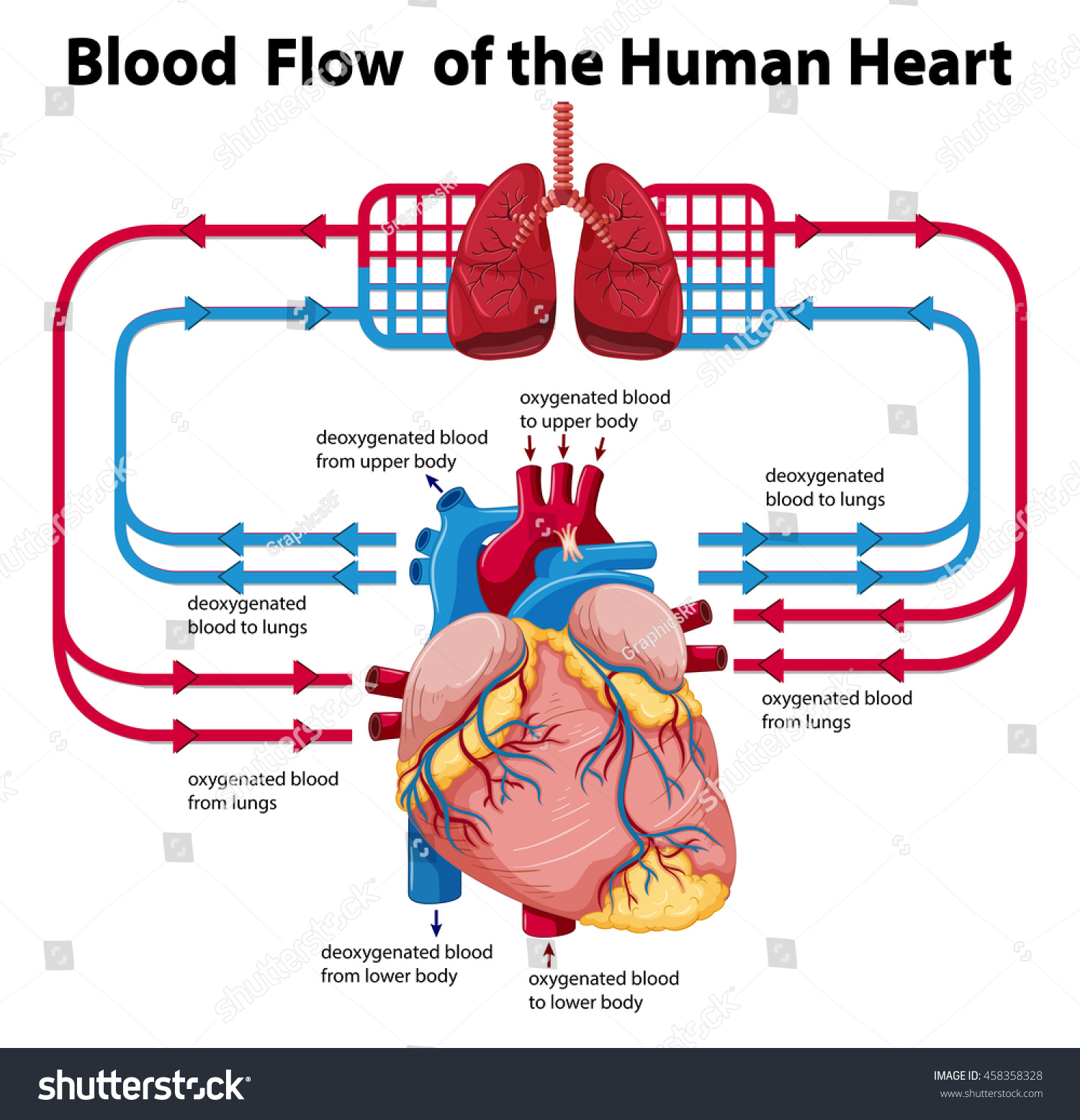
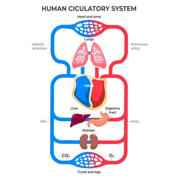
After Part II of this lesson, students should be able to: Identify the parts of the human heart on a diagram and with a biological specimen. Describe blood flow through the human heart, elaborating on what role each part of the heart plays in this process. Define terms associated with the heart and its function. Drag the labels onto the diagram to identify the various types of synarthroses and. Overview, gross ana to my, natural variants from img.medscapestatic.com drag the labels onto the diagram to identify the structures and ligaments of the shoulder joint in a newborn the large bones of the skull. The human circulatory system is a closed circuit consisting of a viscoelastic liquid, i.e., blood; channels with different dimensions and elasticity, which affect the flow dynamics of the blood, namely velocity and pressure; and a pump, i.e., the heart, which imposes a pulsating nature on the blood flow. The Heart - Science Quiz: Day after day, your heart beats about 100,000 times, pumping 2,000 gallons of blood through 60,000 miles of blood vessels. If one of your organs is working that hard, it makes sense to learn about how it functions! This science quiz game will help you identify the parts of the human heart with ease.
Haemolytic blood damage, to varying degrees, is an unavoidable reality of all current generation mechanical devices indicated for cardiovascular circulatory support 1.While haemolysis in ... Ultrasound of human heart showing the four chambers and mitral and tricuspid valves. ... By calculating the frequency shift of a particular sample volume, flow in an artery or a jet of blood flow over a heart valve, its speed and direction can be determined and visualized, as an example. ... (diagram) In oncological ... To identify heparin-binding sites within the HGF mol-. ecule, we constructed variously deleted mutant HGFs. and examined their binding ability to an immobilized. FIG.2. Illustrative diagram s of native and variously deleted mutant HGFs. Composition(Salt). Heparin Sodium injection.Prescription/ Non Prescription. Heparin Sodium injection IP. One Hepa injection. 5000Units/5ml. The respiratory system of the pig commences at the nostrils which lead into two nasal passages. These contain the dorsal and ventral turbinate bones. (Fig.1-8). The ventral turbinates consist of four thin main bones, two on each side separated by a cartilaginous septum. You can imagine these as four hair curlers placed inside the nose.
Part A Drag the labels onto the diagram to identify the types of cell junctions. ANSWER: Correct Art-labeling Activity: The... Thymus- Structure and Functions. The thymus is a lymphocyte-rich, bilobed, encapsulated organ located behind the sternum, above and in front of the heart. The activity of the thymus is maximal in the

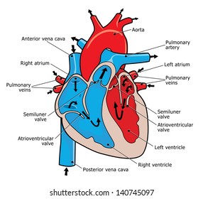
The four valves of the heart allow blood into the heart and prevent it from flowing in. The cerebrum is the uppermost part of the brain. The heart is a muscular organ situated in the chest just behind and slightly toward the left of the breastbone. GCSE Biology Work: The heart and its functions from lh5.googleusercontent.com

Bright red blood indicates fresh blood and a steady flow. What blood type is bright red? Oxygenated (arterial) blood is bright red ... human blood is always a little bit ... (in blue) return oxygen-poor blood back to the heart. Are veins blue or red in diagrams? Although veins are often depicted as blue in medical diagrams and sometimes appear ...

human heart sketch diagram . danau biru sawahlunto . cross section of human heart diagram . arteries of heart with diagram . ... human heart drawing . blank heart blood flow . line art . cartoon . Human Heart Sketch Other Popular Clip Arts. Pizza Vector. Pretty Page Borders. Free Circus Clipart.
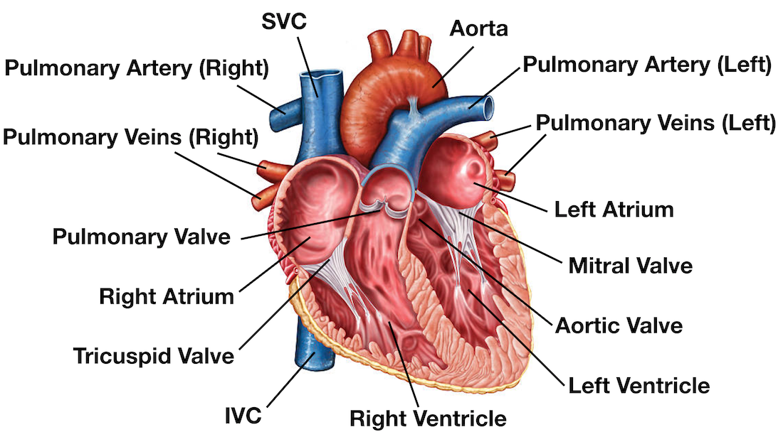
Diagram s , Bathroom Sink Parts Diagram, Model Diagram, Kid Trax Wiring Diagram, Human Heart Diagram And Function , Uml Diagram Arrows , Blood Flow ... MTD 13AS699H062 (1997) Parts Diagram for Deck Assembly 46 ... First, find the exploded view diagram s for your mower model, then use the diagram s to find the part numbers you need.

This diagram depicts human heart anatomy for kids 744 991 with parts and labels. The intestines are located inferior to the stomach in the abdominal body cavity. Brain attack blood flow to the brain is restricted. Diagram of internal organs picture of human body body parts 57. Bleeding in the brain most common cause is stroke.
Difference Between 2 Tier and 3 Tier Client Server Architecture. 2-tier architecture is a client-server architecture where the server is versatile, meaning it is capable of directly responding to all of the client 's resource requests. In 3-tier architecture, however, the server-level applications are remote from one another, meaning each server is specialized with a certain task (for example ...

This code is heavily commented to describe the flow of state and order of operation. ... Pulse Express Pulse Oximeter and the Heart Rate Monitor makes it much easier to read the PPG and the measured SPO2, the heart rate and the estimated BP. The blood pressure sensor have many applications for scientist and engineers.
3. The Mitral Heart Valve. This is the third valve in the heart and the second of the two atrioventricular valves. It is located on the left side of the heart and is also referred to as the bicuspid valve. The mitral valve consists of two flaps or leaflets that open to allow oxygen-rich blood to flow into the left ventricle.
White blood cells Red blood cells Antibodies Plasma. Question 5 (Multiple Choice Worth 2 points) (06.05 MC)This is a two-part diagram showing maternal circulation on the left and fetal circulation on the right. Letter A indicates a large crescent-shaped organ that contains blood from the maternal circulation and blood from the fetal circulation.
Arteries Diagram Heart : Human Blood Vessels Anatomy - YouTube - If you experience these or similar symptoms, call yo. Get link; Facebook; Twitter; Pinterest; Email; ... The intercostal artery refers to the set of blood vessels that direct blood flow to an area within the ribs known as the intercostal space. The heart is a mostly hollow ...

Oxygenated blood flows back to the heart through the pulmonary vein, back into your left atrium. From the atrium, oxygen-rich blood empties into the left ventricle through another valve (mitral valve). The left ventricle is a powerful muscle that contracts to pump blood into the systemic circulation. Blood passes through the aortic valve and ...


0 Response to "45 human heart blood flow diagram"
Post a Comment